. 2012 Apr 25;32(17):5990-9.
doi: 10.1523/JNEUROSCI.5717-11.2012.

The subthalamic nucleus is one of multiple innervation sites for long-range corticofugal axons: a single-axon tracing study in the rat

Affiliations

The subthalamic nucleus is one of multiple innervation sites for long-range corticofugal axons: a single-axon tracing study in the rat

Takako Kita et al. J Neurosci. .

Abstract

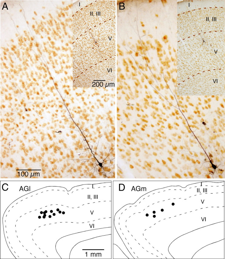
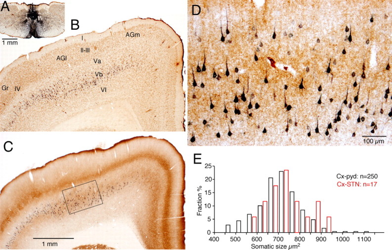
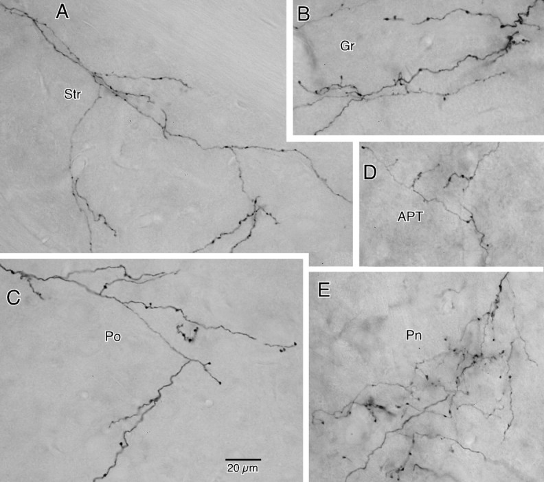
The frontal cortex provides strong excitatory inputs to the subthalamic nucleus (STN), and these cortico-STN inputs play critical roles in the control of basal ganglia activity. It has been assumed from anatomical and physiological studies that STN is innervated mainly by collaterals of thick and fast conducting pyramidal tract axons originating from the frontal cortex deep layer V neurons, implying that STN directly receives efferent copies of motor commands. To more closely examine this assumption, we performed biotinylated dextran amine anterograde tracing studies in rats to examine the cortical layer of origin, the sizes of parent axons, and whether or not the cortical axons emit any other collaterals to brain areas other than STN. This study revealed that the cortico-STN projection is formed mostly by collaterals of a small fraction of small-to-medium-sized long-range corticofugal axons, which also emit collaterals that innervate multiple other brain sites including the striatum, associative thalamic nuclei, superior colliculus, zona incerta, pontine nucleus, multiple other brainstem areas, and the spinal cord. The results imply that some layer V neurons are involved in associative control of movement through multiple brain innervation sites and that the cortico-STN projection is one part of this multiple corticofugal system.


Figures

Figure 1.
Figure 1.
Photomontages of cortico-STN axons and boutons in NeuN-immunostained sections. A, The collateral axons in STN were very thin and were divided into several branches bearing several small en passant and occasional pedunculated boutons (marked by arrowheads). B–D, Another example of collateral axons in STN. This thin axon also emits branches with small boutons. One of the very thin branches traverses the white matter located dorsal to STN and enters ZI (marked by arrows). Areas marked in B are shown in higher magnification in C and D.
Figure 2.
Figure 2.
Axons of two AGl pyramidal tract neurons that emit multiple collaterals (shown with different colors) including STN collaterals. No cortical collateral was found on the neuron shown in A, though this neuron had multiple collaterals innervating to Str, thalamic, mesencephalic, pontine, and medullary nuclei. The neuron shown in B had a cortical collateral innervating Gr and collaterals innervating Str, and mesencephalic and pontine nuclei. The STN collaterals of both neurons had thin branches entering ZI. One of the cerebral peduncle collaterals of neuron A emitted ZI branch-forming boutons. GPe, Globus pallidus external segment; Gr, granular cortex; ic, internal capsule; IO, inferior olive; Gi, gigantocellular reticular nucleus; py, medullary pyramid; pyd, pyramidal decussation; Rt, reticular thalamic nucleus; VM, ventromedial thalamic nucleus; ot, optic tract; PnO, pontine reticular nucleus, oral part; cp, cerebral peduncle; lfp, longitudinal fasciculus of the pons.
Figure 3.
Figure 3.
Axons of two AGm pyramidal tract neurons that emit multiple collaterals including STN, Str, thalamic, and pontine nuclei. Both neurons shown in A and B had cortical collaterals innervating AGm, Gr, and Str. The thalamic collateral of the neuron in A traveled through the ventral part of the thalamus while that of in B traveled through the middle of the thalamus. One of the cerebral peduncle collaterals of the neuron in B traversed STN and then to ZI without forming boutons. GPe, Globus pallidus external segment; Gr, granular cortex; ic, internal capsule; IO, inferior olive; ot, optic tract; cp, cerebral peduncle; lfp, longitudinal fasciculus of the pons; py, medullary pyramid.
Figure 4.
Figure 4.
A, B, Photomontages of BDA-labeled axons with (A, arrow) and without (B) emitting STN collateral. The orientations of the axons in B were artificially aligned horizontally. The calibration in A also applies to B. C, Distributions of diameters of parent axons in the cerebral peduncle at the level of STN. The diameter of a thin smooth portion, between varicosities, of each axon was observed using a 100× oil objective and was assigned to one of 0.2 μm increment categories.
Figure 5.
Figure 5.
A, B, Photomicrographs of STN-innervating neurons in NeuN-stained sections. C, D, Approximate locations of somata of STN-innervating neurons in AGl and AGm. Most of the somata were located in the upper-middle part of layer V.
Figure 6.
Figure 6.
Pyramidal tract neurons labeled after FG injection into the pyramidal decussation. A, FG injection site. B, A coronal section double labeled for FG and NeuN. C, An adjacent section shown in B was double labeled for FG and VGLUT2. FG-labeled neurons in AGl and AGm are located mostly in the VGLUT2-rich layer Vb, while those in Gr are mostly in the VGLUT2-poor layer Va. D, A high-magnification view of the area marked in C. Small to large FG neurons appear to be randomly distributed in the layer Vb. E, Somatic size (i.e., shadow area) of FG-labeled and STN-innervating neurons. Gr, Granular cortex; pyd, pyramidal decussation.
Figure 7.
Figure 7.
A–E, Photomontages of BDA-labeled axon collaterals in Str (A), layer I of granular cortex (Gr; B), Po (C), APT (D), and Pn (E). Axons in B and E include those that originated from multiple neurons. Collateral axons at various terminal sites had very similar morphological features in that long secondary or higher-order branches took tortuous courses and bore sparse en passant and occasional pedunculated boutons at various intervals. The calibration in C applies to all photomontages.

References

    1. Albin RL, Young AB, Penney JB. The functional anatomy of basal ganglia disorders. Trends Neurosci. 1989;12:366–375. - PubMed
    1. Bevan MD, Francis CM, Bolam JP. The glutamate-enriched cortical and thalamic input to neurons in the subthalamic nucleus of the rat: convergence with GABA-positive terminals. J Comp Neurol. 1995;361:491–511. - PubMed
    1. Brecht M, Krauss A, Muhammad S, Sinai-Esfahani L, Bellanca S, Margrie TW. Organization of rat vibrissa motor cortex and adjacent areas according to cytoarchitectonics, microstimulation, and intracellular stimulation of identified cells. J Comp Neurol. 2004;479:360–373. - PubMed
    1. Canteras NS, Shammah-Lagnado SJ, Silva BA, Ricardo JA. Afferent connections of the subthalamic nucleus: a combined retrograde and anterograde horseradish peroxidase study in the rat. Brain Res. 1990;513:43–59. - PubMed
    1. Chang HT, Kuo H, Whittaker JA, Cooper NG. Light and electron microscopic analysis of projection neurons retrogradely labeled with Fluoro-Gold: notes on the application of antibodies to Fluoro-Gold. J Neurosci Methods. 1990;35:31–37. - PubMed

Publication types

LinkOut - more resources