

Multiphoton microscopy in the evaluation of human bladder biopsies
- PMID: 22540300
- PMCID: PMC3640548
- DOI: 10.5858/arpa.2011-0147-OA
Multiphoton microscopy in the evaluation of human bladder biopsies
Abstract
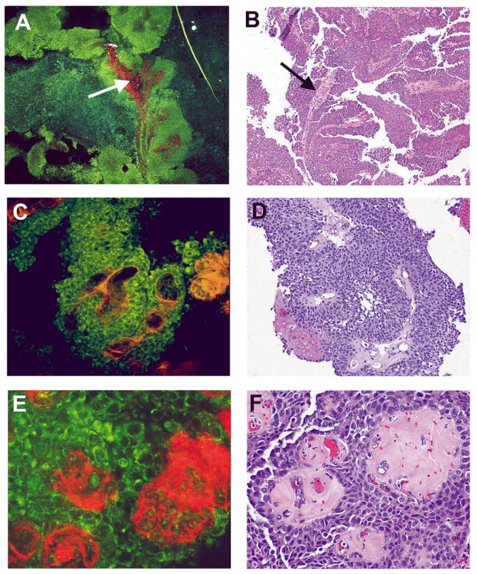
Context: Multiphoton microscopy (MPM) is a nonlinear imaging approach, providing cellular and subcellular details from fresh (unprocessed) tissue by exciting intrinsic tissue emissions. With miniaturization and substantially decreased cost on the horizon, MPM is an emerging imaging technique with many potential clinical applications.
Objectives: To assess the imaging ability and diagnostic accuracy of MPM for human bladder biopsies.
Design: Seventy-seven fresh bladder biopsies were imaged by MPM and subsequently submitted for routine surgical pathology diagnosis. Twelve cases were excluded because of extensive cautery artifact that prohibited definitive diagnosis. Comparison was made between MPM imaging and gold standard sections for each specimen stained with hematoxylin-eosin.
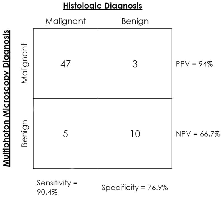
Results: In 57 of 65 cases (88%), accurate MPM diagnoses (benign or neoplastic) were given based on the architecture and/or the cytologic grade. The sensitivity and specificity of MPM in our study were 90.4% and 76.9%, respectively. A positive (neoplastic) diagnosis on MPM had a high predictive value (94%), and negative (benign) diagnoses were sustained on histopathology in two-thirds of cases. Architecture (papillary versus flat) was correctly determined in 56 of 65 cases (86%), and cytologic grade (benign/low grade versus high grade) was assigned correctly in 38 of 56 cases (68%).
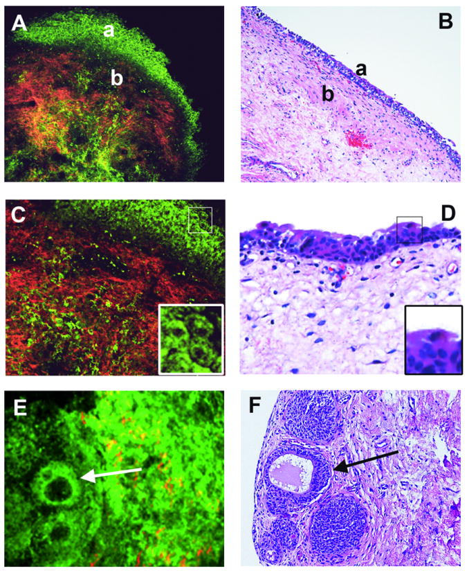
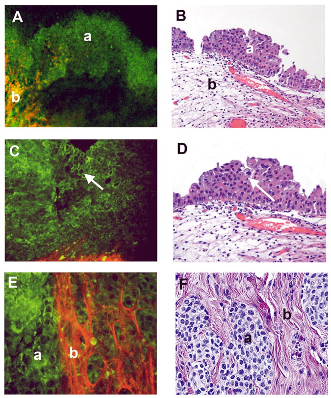
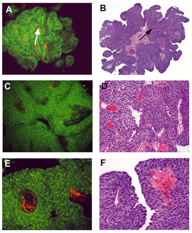
Conclusions: The MPM images alone provided sufficient detail to classify most lesions as either benign or neoplastic using the same basic diagnostic criteria as histopathology (architecture and cytologic grade). Future developments in MPM technology may provide urologists and pathologists with additional screening and diagnostic tools for early detection of bladder cancer. Additional applications of such emerging technologies warrant exploration.
Figures
Similar articles
-
Multiphoton microscopy: a potential intraoperative tool for the detection of carcinoma in situ in human bladder.Arch Pathol Lab Med. 2015 Jun;139(6):796-804. doi: 10.5858/arpa.2014-0076-OA. Arch Pathol Lab Med. 2015. PMID: 26030249
-
Multiphoton tomographic imaging: a potential optical biopsy tool for detecting gastrointestinal inflammation and neoplasia.Cancer Prev Res (Phila). 2012 Nov;5(11):1280-90. doi: 10.1158/1940-6207.CAPR-12-0132. Epub 2012 Sep 7. Cancer Prev Res (Phila). 2012. PMID: 22961775 Free PMC article.
-
Real-time optical diagnosis for surgical margin in low rectal cancer using multiphoton microscopy.Surg Endosc. 2014 Jan;28(1):36-41. doi: 10.1007/s00464-013-3153-7. Epub 2013 Sep 4. Surg Endosc. 2014. PMID: 24002915
-
Multiphoton microscopy in surgical oncology- a systematic review and guide for clinical translatability.Surg Oncol. 2019 Dec;31:119-131. doi: 10.1016/j.suronc.2019.10.011. Epub 2019 Oct 16. Surg Oncol. 2019. PMID: 31654957
-
Frontiers in Intravital Multiphoton Microscopy of Cancer.Cancer Rep (Hoboken). 2020 Feb;3(1):e1192. doi: 10.1002/cnr2.1192. Epub 2019 Jun 20. Cancer Rep (Hoboken). 2020. PMID: 32368722 Free PMC article. Review.
Cited by
-
Optical biomarkers of serous and mucinous human ovarian tumor assessed with nonlinear optics microscopies.PLoS One. 2012;7(10):e47007. doi: 10.1371/journal.pone.0047007. Epub 2012 Oct 8. PLoS One. 2012. PMID: 23056557 Free PMC article.
-
Multiphoton microscopy to identify and characterize the transition zone in a mouse model of Hirschsprung disease.J Pediatr Surg. 2013 Jun;48(6):1288-93. doi: 10.1016/j.jpedsurg.2013.03.025. J Pediatr Surg. 2013. PMID: 23845620 Free PMC article.
-
Enabling Multiphoton and Second Harmonic Generation Imaging in Paraffin-Embedded and Histologically Stained Sections.Tissue Eng Part C Methods. 2016 Jun;22(6):517-23. doi: 10.1089/ten.TEC.2016.0071. Epub 2016 Apr 25. Tissue Eng Part C Methods. 2016. PMID: 27018844 Free PMC article.
-
Emerging endoscopic imaging technologies for bladder cancer detection.Curr Urol Rep. 2014 May;15(5):406. doi: 10.1007/s11934-014-0406-5. Curr Urol Rep. 2014. PMID: 24658832 Free PMC article. Review.
-
[Supplementary optical techniques for the detection of nonmuscle invasive bladder cancer].Urologe A. 2018 Feb;57(2):139-147. doi: 10.1007/s00120-017-0539-5. Urologe A. 2018. PMID: 29110046 Review. German.
References
-
- Jemal A, Siegel R, Xu J, Ward E. Cancer statistics, 2010. CA Cancer J Clin. 2010;60:277–300. - PubMed
-
- Epstein J, Amin M, Reuter V. Biopsy interpretation of the bladder. 2. Philadelphia, PA: Lippincott Williams & Wilkins; 2010.
-
- Messing E. Urothelial Tumors of the Bladder. In: Wein AJ, editor. Wein: Campbell-Walsh Urology. 9. Vol. 3. PA: Saunders Elsevier; 2007.
-
- Epstein J, Amin M, Reuter V, Mostofi F. The World Health Organization/International Society of Urological Pathology Consensus Classification of Urothelial (Transitional Cell) Neoplasms of the Urinary Bladder. The American Journal of Surgical Pathology; The Bladder Consensus Conference Committee; 1998. pp. 1435–1448. - PubMed
-
- Jones S, Campbell S. Non–Muscle-Invasive Bladder Cancer (Ta, T1, and CIS) 9. Vol. 3. PA: Saunders Elsevier; 2007.
Publication types
MeSH terms
Grants and funding
LinkOut - more resources
Full Text Sources
Other Literature Sources
Medical