Review
. 2012:197:101-21.
doi: 10.1016/B978-0-444-54299-1.00006-6.

Neurological phenotypes for Down syndrome across the life span

Affiliations
Review

Neurological phenotypes for Down syndrome across the life span

Ira T Lott. Prog Brain Res. 2012.

Abstract

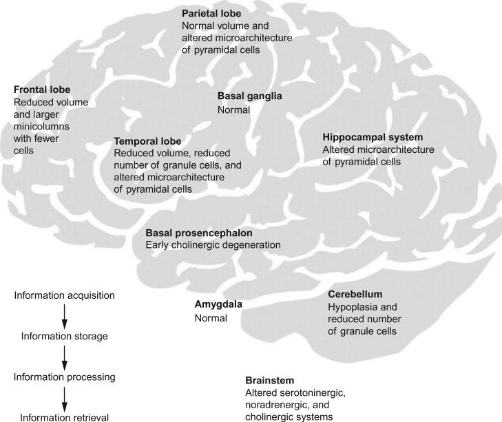
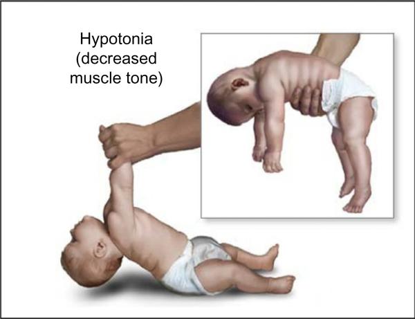
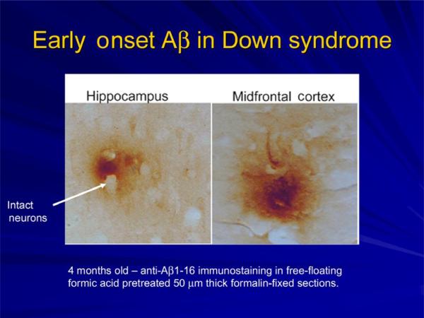
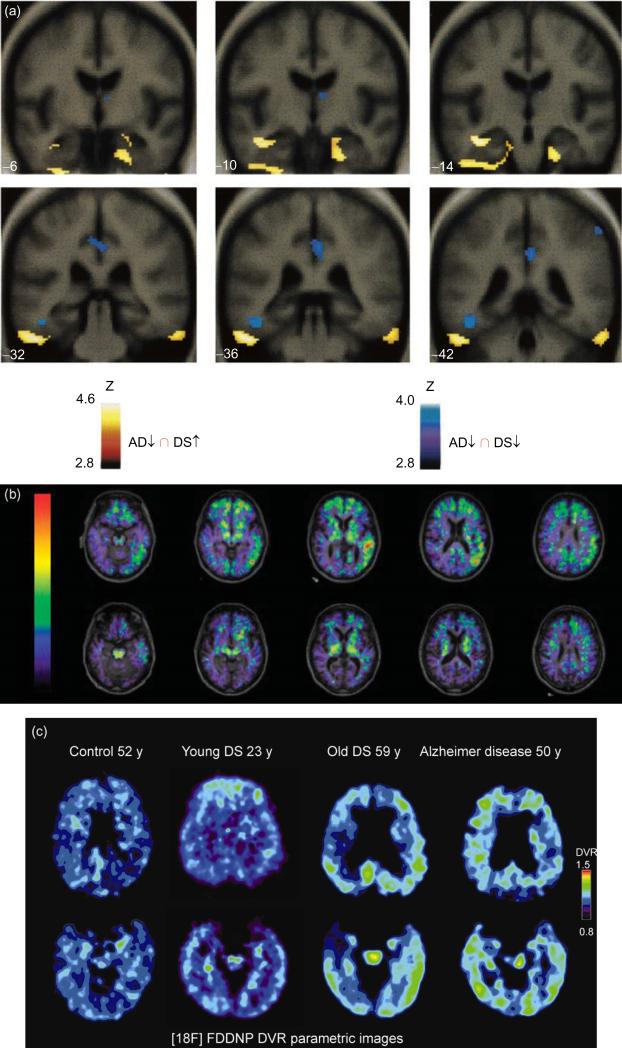
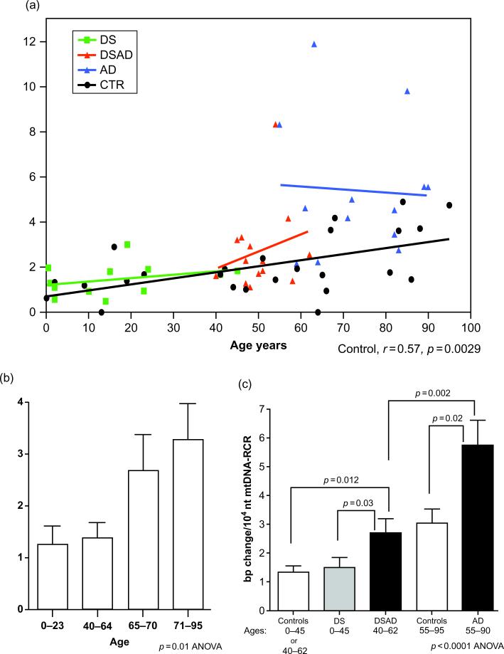
This chapter reviews the neurological phenotype of Down syndrome (DS) in early development, childhood, and aging. Neuroanatomic abnormalities in DS are manifested as aberrations in gross brain structure as well as characteristic microdysgenetic changes. As the result of these morphological abnormalities, brain circuitry is impaired. While an intellectual disability is ubiquitous in DS, there is a wide range of variation in cognitive performance and a growing understanding between aberrant brain circuitry and the cognitive phenotype. Hypotonia is most marked at birth, affecting gait and ligamentous laxity. Seizures are bimodal in presentation with infantile spasms common in infancy and generalized seizures associated with cognitive decline observed in later years. While all individuals have the characteristic neuropathology of Alzheimer's disease (AD) by age 40 years, the prevalence of dementia is not universal. The tendency to develop AD is related, in part, to several genes on chromosome 21 that are overexpressed in DS. Intraneuronal accumulation of β-amyloid appears to trigger a cascade of neurodegeneration resulting in the neuropathological and clinical manifestations of dementia. Functional brain imaging has elucidated the temporal sequence of amyloid deposition and glucose metabolic rate in the development of dementia in DS. Mitochondrial abnormalities contribute to oxidative stress which is part of AD pathogenesis in DS as well as AD in the general population. A variety of medical comorbidities threaten cognitive performance including sleep apnea, abnormalities in thyroid metabolism, and behavioral disturbances. Mouse models for DS are providing a platform for the formulation of clinical trials with intervention targeted to synaptic plasticity, brain biochemistry, and morphological brain alterations.


Figures

Fig. 1
Fig. 1
The learning circuit in DS. Structures involved in information acquisition, processing, or storage are affected in DS. The figure depicts some of the described changes that may lead to cognitive dysfunction in individuals with DS. (Reprinted from Lancet Neurology, Lott IT and Dierssen M, Cognitive deficits and associated neurological complications in individuals with Down's syndrome, 2010, June 9(6):623–33 with permission from Elsevier).
Fig. 2
Fig. 2
Hypotonia in Down syndrome. Note the head lag upon pull to sitting and the inability to support posture in ventral suspension.
Fig. 3
Fig. 3
Aβ deposition in brain of an infant with Down syndrome. (Image provided by Dr. Elizabeth Head, Sanders-Brown Center on Aging, University of Kentucky, Lexington, KY).
Fig. 4
Fig. 4
(a) FDG imaging in Down syndrome (DS) (Reprinted from Neurology, Haier RJ, et al., Temporal cortex hypermetabolism in Down syndrome prior to the onset of dementia, 2003, Dec 23;61(12):1673–1679, with permission from Wolter Kluwer Health). Results of SPM'99 conjunction analyses. Blue areas show where glucose metabolic rate (GMR) is lower in subjects with Alzheimer's disease (AD) compared with matched controls and where GMR is lower in nondemented DS subjects compared with matched controls. Yellow areas show the conjunction of where GMR is higher in the nondemented DS group compared with matched controls and where GMR is lower in the AD subjects compared with their matched controls. Note: The yellow areas are in the inferior temporal/entorhinal cortex; blue areas are in the posterior cingulate and left fusiform gyrus. Statistical results are shown on MRI templates for six coronal slices (-6-42), which show a large section of inferiotemporal/entorhinal cortex. Talairach and Tournoux atlas coordinates of all findings (p<0.001). (b) PiB amyloid staining for dementia in DS (Reprinted from Archives of Neurology, Landt J et al., Using positron emission tomography and carbon 11-labeled Pittsburgh compound B to image brain fibrillar β-amyloid in adults with Down syndrome: Safety, acceptability, and feasibility, 2011, July; 68 (7):890–896 with permission from American Medical Association). Fused carbon 11-labeled Pittsburgh Compound B nondisplaceable binding potential and magnetic resonance images for a subject with DS and AD (top) and a control without DS (bottom). (c) FDDNP imaging in DS (Reprinted from Archives of Neurology, Nelson LD et al., Positron emission tomogrpahy of brain β-amyloid and tau levels in adults with Down syndrome, 2011, June; 68(6):768–774 with permission from American Medical Association). 18F-FDDNP PET imaging in control, young DS, old DS, and AD showing binding characteristics.
Fig. 5
Fig. 5
mtDNA Regulatory Control Region (RCR) mutation frequency in the frontal cortices of control, DS, DSAD, and AD patients. (a) The correlation of mtDNA RCR mutation frequency with aging in all four groups. (b) The comparison of mtDNA RCR mutation frequency in four different age groups: 0–23, 40–64, 65–70, and 71–95years. (c) The comparison of mtDNA RCR mutation frequencies of DSAD and AD brains to age-matched controls and DS brains. Because the control groups for DS (ages 0–40) and DSAD (ages 40–62) were not significantly different (see panel b), DS and DSAD control groups were grouped together. (Reprinted from Journal of Alzheimer's Disease, Coskun P et al., Mitochondrial dysfunction and the etiology of Alzheimer's disease and Down syndrome dementia, 2010:20, S293–S310, with permission from IOS press).

References

    1. Agiovlasitis S, McCubbin JA, Yun J, Pavol MJ, Widrick JJ. Economy and preferred speed of walking in adults with and without Down syndrome. Adapted Physical Activity Quarterly. 2009;26:118–130. - PubMed
    1. Amaral DG, Schumann CM, Nordahl CW. Neuroanatomy of autism. Trends in Neurosciences. 2008;31:137–145. - PubMed
    1. Andrade DM. Genetic basis in epilepsies caused by malformations of cortical development and in those with structurally normal brain. Human Genetics. 2009;126:173–193. - PubMed
    1. Andreou G, Galanopoulou C, Gourgoulianis K, Karapetsas A, Molyvdas P. Cognitive status in Down syndrome individuals with sleep disordered breathing deficits (SDB). Brain and Cognition. 2002;50:145–149. - PubMed
    1. Andriolo RB, El Dib RP, Ramos L, Atallah AN, da Silva EM. Aerobic exercise training programmes for improving physical and psychosocial health in adults with Down syndrome. Cochrane Database of Systematic Reviews. 2010:CD005176. - PubMed

Publication types

Substances