. 2012 Jun 1;11(6):537-49.
doi: 10.1016/j.dnarep.2012.04.003. Epub 2012 Apr 26.

RECQ1 plays a distinct role in cellular response to oxidative DNA damage

Affiliations

RECQ1 plays a distinct role in cellular response to oxidative DNA damage

Sudha Sharma et al. DNA Repair (Amst). .

Abstract

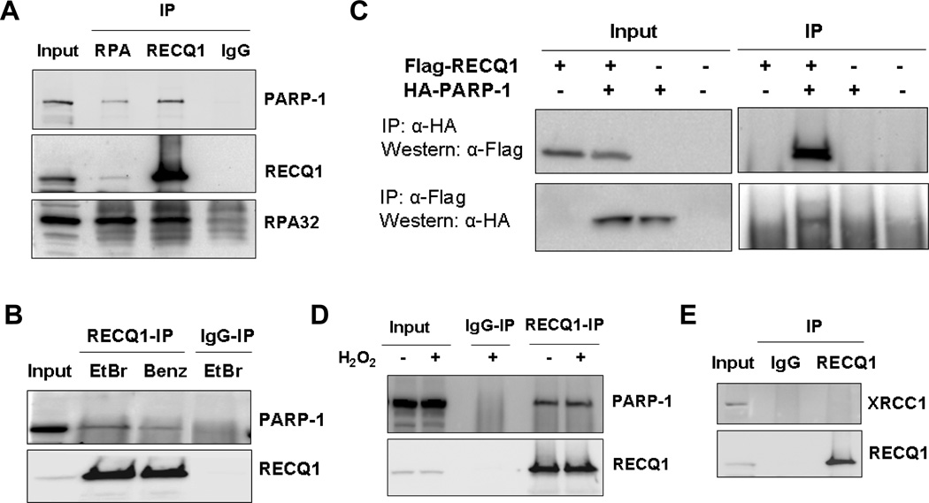
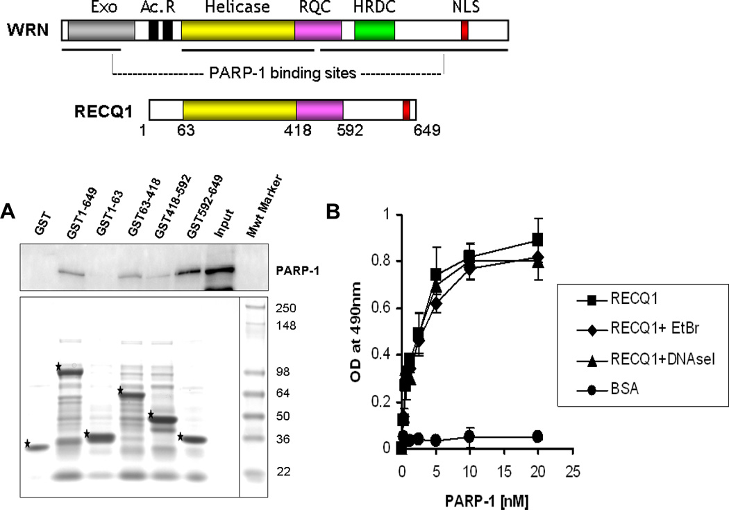
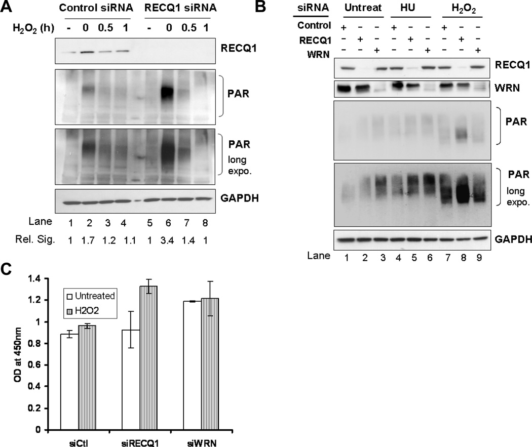
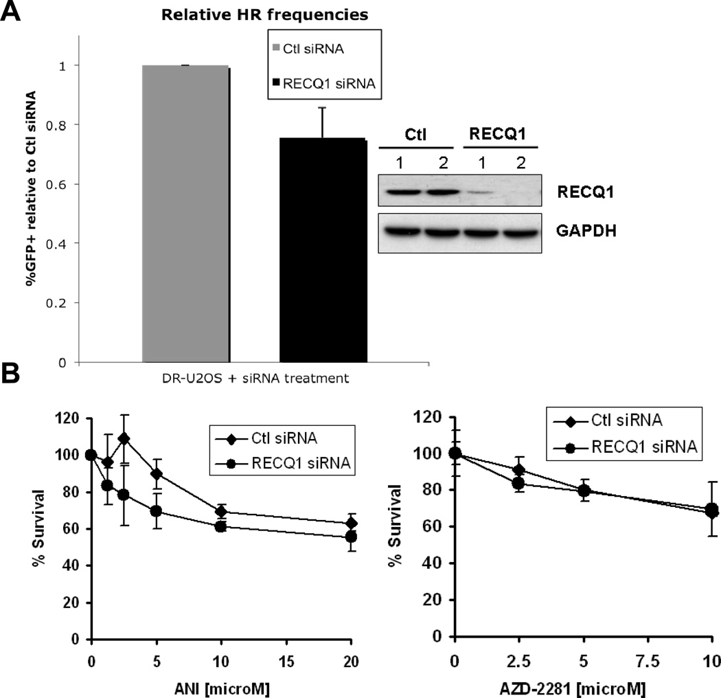
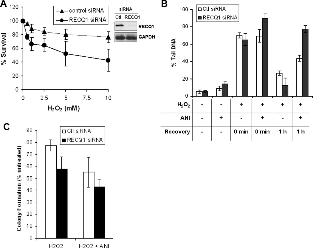
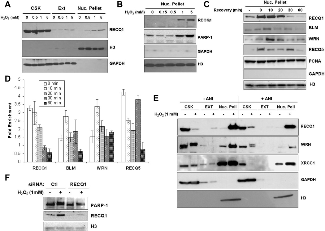
RECQ1 is the most abundant RecQ homolog in humans but its functions have remained mostly elusive. Biochemically, RECQ1 displays distinct substrate specificities from WRN and BLM, indicating that these RecQ helicases likely perform non-overlapping functions. Our earlier work demonstrated that RECQ1-deficient cells display spontaneous genomic instability. We have obtained key evidence suggesting a unique role of RECQ1 in repair of oxidative DNA damage. We show that similar to WRN, RECQ1 associates with PARP-1 in nuclear extracts and exhibits direct protein interaction in vitro. Deficiency in WRN or BLM helicases have been shown to result in reduced homologous recombination and hyperactivation of PARP under basal condition. However, RECQ1-deficiency did not lead to PARP activation in undamaged cells and nor did it result in reduction in homologous recombination repair. In stark contrast to what is seen in WRN-deficiency, RECQ1-deficient cells hyperactivate PARP in a specific response to H₂O₂treatment. RECQ1-deficient cells are more sensitive to oxidative DNA damage and exposure to oxidative stress results in a rapid and reversible recruitment of RECQ1 to chromatin. Chromatin localization of RECQ1 precedes WRN helicase, which has been shown to function in oxidative DNA damage repair. However, oxidative DNA damage-induced chromatin recruitment of these RecQ helicases is independent of PARP activity. As other RecQ helicases are known to interact with PARP-1, this study provides a paradigm to delineate specialized and redundant functions of RecQ homologs in repair of oxidative DNA damage.


Conflict of interest statement

Conflict of Interests

The authors declare that there are no conflicts of interest

Figures

Figure 1
Figure 1. RECQ1 interacts with PARP-1 in vivo
A, Co-immunoprecipitation analysis of RECQ1 interaction with PARP-1 using HeLa nuclear extracts. Immunoprecipitations (IP) with antibodies specific for RECQ1, RECQ1 interacting protein RPA, and preimmune IgG are indicated. Eluted proteins in immunoprecipitate were analyzed by Western blotting and are indicated. Reciprocal co-IPs of RECQ1 and RPA also contained PARP-1. B, Association of RECQ1 and PARP-1 is not mediated via DNA. RECQ1 antibody co-precipitated RECQ1 and PARP-1 in the presence of EtBr or using benzonase-treated extract in IP reaction. C, Reciprocal co-IP of ectopically expressed RECQ1 and PARP-1. HeLa cells were untransfected or transiently transfected with Flag-RECQ1, HA-PARP-1 or both. Lysates of these cells were used for IP using anti-HA or anti-Flag antibody and analyzed by Western blotting as indicated. D. Association between RECQ1-PARP-1 is not modulated by H2O2-induced DNA damage. HeLa cells were untreated or treated with H2O2 (1 mM, 20 min) and harvested immediately. IP with anti-RECQ1 antibody was followed by Western analyses using anti-PARP-1 and anti-RECQ1 antibodies. E. XRCC1 does not co-IP with RECQ1. RECQ1 IP was subjected to Western analysis using anti-XRCC1 or anti-RECQ1 antibodies. In all experiments, input corresponds to 5% of total protein used in IP reactions.
Figure 2
Figure 2. A direct physical interaction between RECQ1-PARP-1 is mediated via the helicase domain and C-terminal region of RECQ1 in vitro
A. Top: A schematic diagram of the full-length WRN and RECQ1 proteins showing conserved domains. RQC, RecQ-conserved domain; HRDC, helicase RnaseD conserved domain; NLS, nuclear localization signal. Also shown are Exonuclease (Exo) domain and Acidic repeats (Ac. R) present in WRN protein. Previously reported PARP-1 binding regions of WRN are indicated by horizontal lines. Bottom: Identification of the PARP-1 interacting region of RECQ1. GST alone or GST-RECQ1 fragments (as indicated) bound to glutathione beads were incubated with HeLa extracts (500 µg) overnight at 4°C. After extensive washings, the bound PARP-1 was eluted with SDS sample buffer and analyzed by Western blot using anti-PARP-1 antibody (upper). Coomassie staining of the eluted proteins was done to test expression of various GST-fusion fragments of RECQ1 (lower). GST-RECQ1 proteins are marked by asterisk. Mwt Marker, protein molecular weight marker. B. Recombinant RECQ1 and PARP-1 proteins interact directly as shown by ELISA. Either BSA or purified recombinant RECQ1 was coated onto microtiter plates. Following blocking with 3% BSA, appropriate wells were incubated with the indicated concentrations of recombinant PARP-1 (0–20 nM) for 1 h at 30°C. Parallel wells contained DNaseI (100 U/ml) or EtBr (50 µg/ml) in the binding step to test for DNA-mediated protein interaction. Following washing, RECQ1 bound PARP-1 was detected by ELISA using anti-PARP-1 antibody. The values represent the mean of three independent experiments performed in duplicate with SD indicated by error bars.
Figure 3
Figure 3. RECQ1-deficient cells exhibit increased activation of PARP upon H2O2 exposure
36 h after transfection with control, RECQ1, or WRN siRNA, HeLa cells were treated with 200 µM H2O2 for 20 min and allowed to recover in complete medium. Lysates were prepared at indicated time points following treatment and PARP activation was determined by the intensity of poly-ADP(ribose) (PAR) signal using Western blot analyses (both short and long exposure of Western blot are shown). Depletion of RECQ1 or WRN in the lysates was verified by Western blot and is indicated. GAPDH signal in the same blot serves as loading control. A. RECQ1 depleted cells show increased PAR formation upon H2O2 treatment as compared to control cells (lane 2 vs. lane 6). Control and RECQ1-depleted cells show comparable level of PAR formation in untreated condition. Total PAR signal on Western blot was quantified using ImageJ and signal intensity relative to untreated is indicated in each case. Rel. Sig., relative signal intensity for PAR. B. Depletion of RECQ1 or WRN leads to distinct cellular response with respect to PARP activation upon H2O2 exposure. PAR signal was detected in lysates prepared from control, RECQ1 or WRN-depleted cells that were either untreated or exposed to 2 mM hydroxyurea (HU) for 16h or 200 µM H2O2 for 20 min. C. PARP activity was measured by a quantitative colorimetric assay using lysates from control, RECQ1 or WRN-depleted cells that were either untreated or treated with 200 µM H2O2 for 20 min. PARP activity was measured by taking OD at 450 nm. Values represent mean of three independent experiments with SD indicated by error bars. Ctl, control.
Figure 4
Figure 4. RECQ1 does not play a direct role in homologous recombination repair of DNA double-strand breaks
A. DR-U2OS cells were transfected with control or RECQ1 siRNA twice. Upon second siRNA transfection (at I-SceI TF), cells were either harvested for Western blot analysis or additionally transfected with pCAGGS (empty vector) or pCABSce (I-SceI expression vector). 48 h after second siRNA transfection (at FACs), cells were harvested for Western blot analysis or analyzed for GFP fluorescence. Restoration of a functional GFP gene through complete homologous recombination (HR) was used to determine HR proficiency. %GFP positive cells were quantitated and relative frequencies were determined by normalizing to control siRNA transfected cells. Mean of relative %GFP positive cells and S.E.M. from four independent experiments are shown. Depletion of RECQ1 at I-SceI TF and at FACs, indicated by 1 and 2, respectively, was verified by Western blotting and is shown. GAPDH is used as loading control. B. RECQ1-depleted cells are not sensitive to PARP inhibitors. Cells transfected with control or RECQ1 siRNA were subjected to continuous exposure to increasing dose of ANI or AZD2281 in complete medium and their survival was measured 72 h later by MTS assay. Percentage of control growth was plotted for each data point, representing the mean ± SD of three independent experiments performed in quadruplicate. Ctl, control.
Figure 5
Figure 5. RECQ1-depleted cells repair H2O2-induced DNA damage in PARP activity-dependent manner
A. RECQ1-depleted cells are sensitive to H2O2 exposure. Cells transfected with control or RECQ1 siRNA were exposed in quadruplicate to increasing doses of H2O2 for 10 min followed by growth in complete medium and their survival was measured 72 h later by MTS assay. Percentage of control growth was plotted for each data point, representing the mean ± SD of three independent experiments. Depletion of RECQ1 is shown by Western blot. GAPDH serves as loading control. B. Total DNA strand breaks in control or RECQ1-depleted cells. Alkaline Comet Assay was used to measure the total DNA strand breaks with or without prior exposure to PARP inhibitor ANI (100 µM, 16 h) in control or RECQ1-depleted cells that were either untreated or allowed to recover for indicated time following treatment with H2O2 (10 µM, 5 min). The total DNA strand breaks were quantitated as % tail DNA representing the mean of three independent experiments with SD indicated by error bars. At least 100 cells were analyzed in each experiment. C. Colony formation of H2O2-treated control or RECQ1-depleted cells grown in the presence or absence of ANI. Cells were incubated with or without ANI (10 µM, 16 h) before treatment with H2O2 (1 mM, 10 min) or left untreated. Following removal of H2O2, cells were grown in the presence or absence of ANI (10 µM) in complete medium and colonies were stained and counted after 14 days. As reported previously, depletion of RECQ1 resulted in reduced colony formation [3]. Results are expressed as the relative percentage of colonies as compared to untreated controls for each siRNA following adjustment for plating efficiency using untreated plates and are the means of duplicate experiments, each performed in triplicate. Ctl, control.
Figure 6
Figure 6. RECQ1 associates with damaged chromatin in PARP activity-independent manner
Sequential extraction of HeLa cells without and with H2O2 treatment. CSK and EXT fractions contain cytoplasmic, cytoskeletal, and soluble nuclear proteins. Insoluble nuclear pellet fractions contain proteins that are tightly bound to chromatin and/or the nuclear matrix. GAPDH and histone H3 serve as loading controls and markers for the fractions containing soluble proteins and chromatin, respectively. Results were documented using different exposures of the Western blots. A. RECQ1 associates with chromatin in a H2O2 dose-dependent manner. HeLa cells were treated with the indicated concentrations of H2O2 for 20 min at 37°C followed by sequential fractionation. B. H2O2 dose-dependent chromatin association of RECQ1 is accompanied by enrichment of PARP-1 on damaged chromatin. Nuclear pellet fractions containing chromatin bound proteins are shown. C. Rapid and reversible association of RECQ1 with damaged chromatin. HeLa cells were treated with 1 mM H2O2 for 20 min at 37° followed by incubation in H2O2-free medium for recovery, and subjected to sequential fractionation at indicated time points. Nuclear pellet fractions show relocalization of RECQ1, WRN, BLM and RecQ5β to damaged chromatin at different time during recovery. Levels of PCNA remain unchanged and serve as loading control. Untr., Untreated. D. Quantitation of chromatin enrichment of RecQ proteins at different time points following recovery from H2O2 (1 mM, 20 min) treatment. Western blots were quantitated using ImageJ. Signals were normalized using Histone (H3) and protein signal in the untreated was used to calculate fold enrichment following H2O2 treatment. Mean of relative fold enrichment and S.E.M. from three independent experiments are shown. E. H2O2-induced chromatin binding of RECQ1 and WRN is independent of PARP activity. HeLa cells grown in the presence or absence of ANI (100 µM) for 24 h, were mock-treated or treated with 1 mM H2O2 for 20 min. Cells were then sequentially fractionated and proteins from each fraction analyzed by Western blotting. The apparent decrease of RECQ1, WRN and XRCC1 proteins in the CSK fraction reflects the decrease of soluble nuclear proteins, which now associate tightly with the chromatin-enriched nuclear pellet. F. RECQ1-depleted cells show H2O2 -induced chromatin enrichment of PARP-1. PARP-1 was detected by Western blot in the chromatin fractions prepared from control or RECQ1-depleted cells that were either untreated or treated with 1 mM H2O2 for 20 min.

References

    1. Sharma S, Doherty KM, Brosh RM., Jr Mechanisms of RecQ helicases in pathways of DNA metabolism and maintenance of genomic stability. Biochem J. 2006;398:319–337. - PMC - PubMed
    1. Kawabe T, Tsuyama N, Kitao S, Nishikawa K, Shimamoto A, Shiratori M, Matsumoto T, Anno K, Sato T, Mitsui Y, Seki M, Enomoto T, Goto M, Ellis NA, Ide T, Furuichi Y, Sugimoto M. Differential regulation of human RecQ family helicases in cell transformation and cell cycle. Oncogene. 2000;19:4764–4772. - PubMed
    1. Sharma S, Brosh RM., Jr Human RECQ1 is a DNA damage responsive protein required for genotoxic stress resistance and suppression of sister chromatid exchanges. PLoS ONE. 2007;2:e1297. - PMC - PubMed
    1. Futami K, Kumagai E, Makino H, Goto H, Takagi M, Shimamoto A, Furuichi Y. Induction of mitotic cell death in cancer cells by small interference RNA suppressing the expression of RecQL1 helicase. Cancer Sci. 2008;99:71–80. - PubMed
    1. Futami K, Kumagai E, Makino H, Sato A, Takagi M, Shimamoto A, Furuichi Y. Anticancer activity of RecQL1 helicase siRNA in mouse xenograft models. Cancer Sci. 2008;99:1227–1236. - PMC - PubMed

Publication types

MeSH terms