. 2012 May 25;46(4):449-60.
doi: 10.1016/j.molcel.2012.03.023. Epub 2012 Apr 26.

Nitric oxide modulates bacterial biofilm formation through a multicomponent cyclic-di-GMP signaling network

Affiliations

Nitric oxide modulates bacterial biofilm formation through a multicomponent cyclic-di-GMP signaling network

Lars Plate et al. Mol Cell. .

Abstract

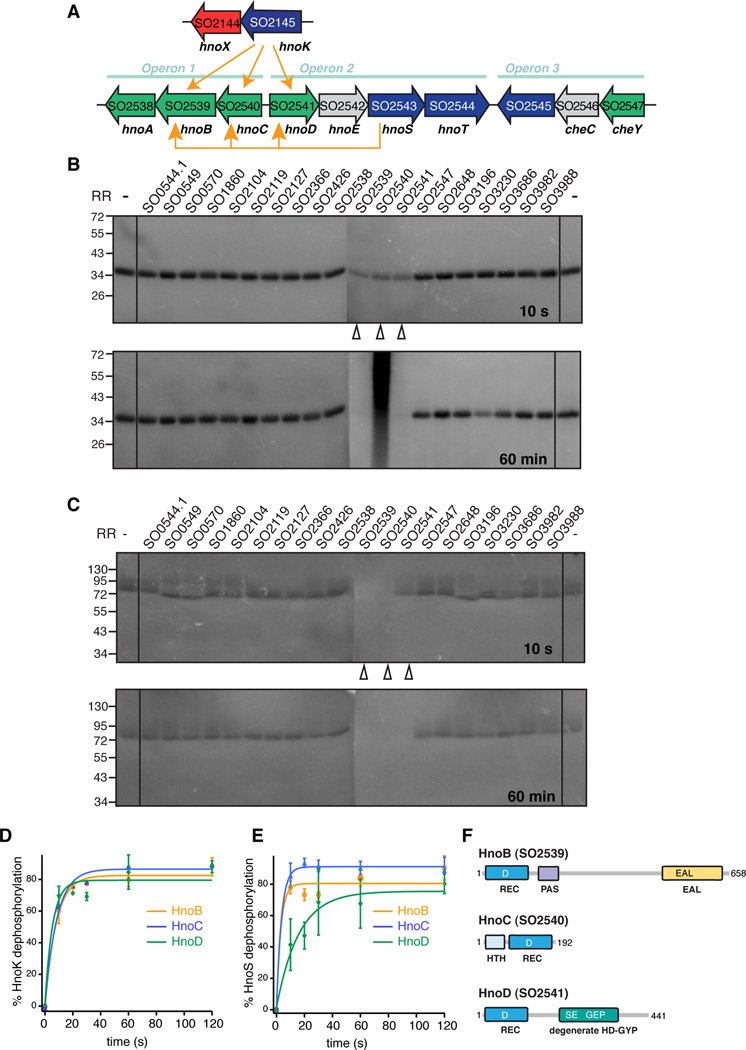
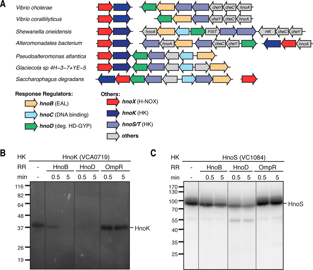
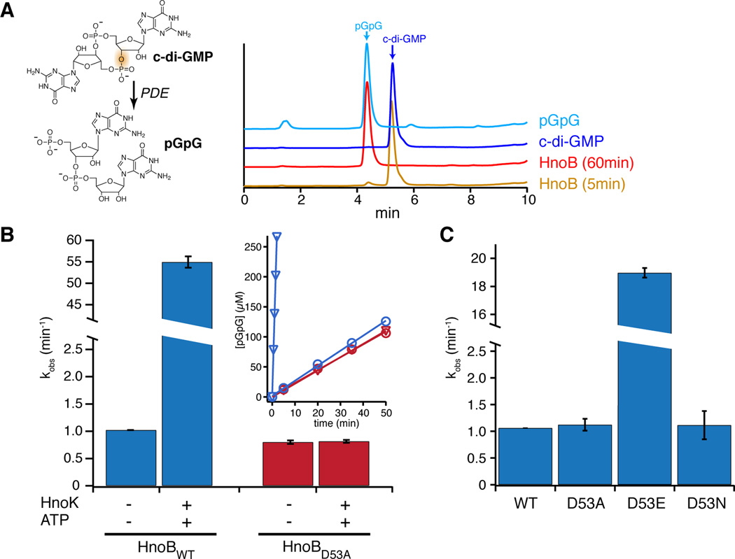
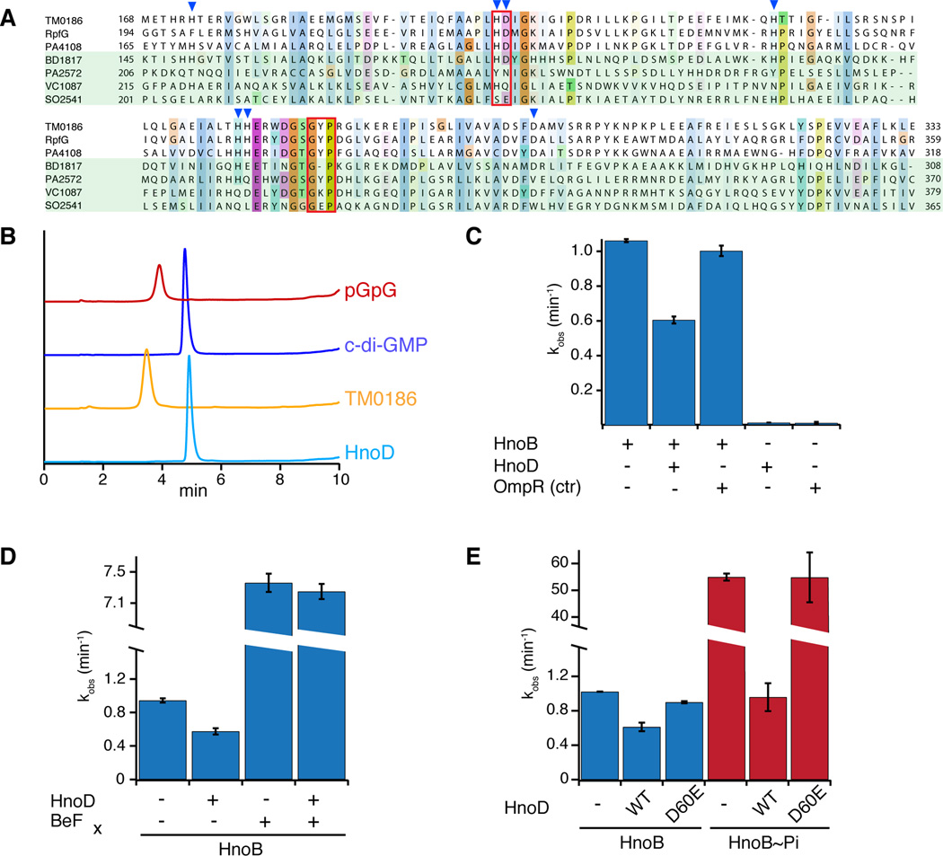
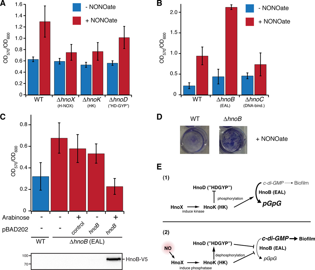
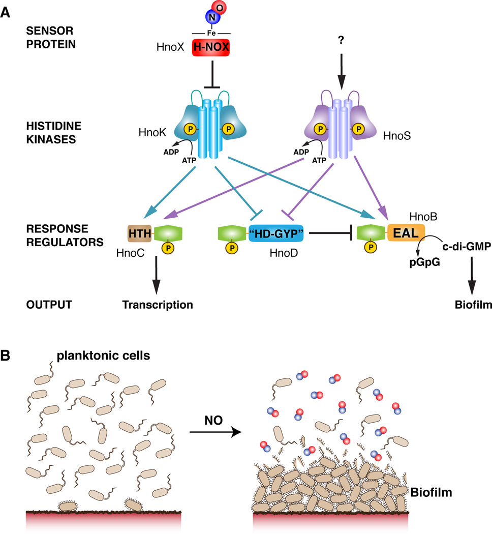
Nitric oxide (NO) signaling in vertebrates is well characterized and involves the heme-nitric oxide/oxygen-binding (H-NOX) domain of soluble guanylate cyclase as a selective NO sensor. In contrast, little is known about the biological role or signaling output of bacterial H-NOX proteins. Here, we describe a molecular pathway for H-NOX signaling in Shewanella oneidensis. NO stimulates biofilm formation by controlling the levels of the bacterial secondary messenger cyclic diguanosine monophosphate (c-di-GMP). Phosphotransfer profiling was used to map the connectivity of a multicomponent signaling network that involves integration from two histidine kinases and branching to three response regulators. A feed-forward loop between response regulators with phosphodiesterase domains and phosphorylation-mediated activation intricately regulated c-di-GMP levels. Phenotypic characterization established a link between NO signaling and biofilm formation. Cellular adhesion may provide a protection mechanism for bacteria against reactive and damaging NO. These results are broadly applicable to H-NOX-mediated NO signaling in bacteria.


Figures

Figure 1
Figure 1. Identification of cognate response regulators to the H-NOX-associated histidine kinase in Shewanella oneidensis
(A) The H-NOX gene (hnoX, SO2144, red) and the H-NOX-associated HK gene (hnoK, SO2145, purple) form an isolated operon in Shewanella oneidensis. The three cognate RR genes of the H-NOX-associated HK hnoB (SO2539), hnoC (SO2540) and hnoD (SO2541) (green) are located in three operons that are rich in two-component signaling proteins. The adjacent HK hnoS (SO2543, purple) had the same three response regulators targets (yellow arrows). (B) Phosphotransfer profiling of the H-NOX-associated HK (HnoK, SO2145) to 20 orphan RRs identified three cognate phosphorylation targets (SO2539, SO2540, SO2541). The HK was pre-phosphorylated with [γ-32P]-ATP and subsequently incubated with an equimolar amount of the respective RR for either 10 s or 60 min. Phosphorylation was detected by visualizing 32P radioactivity after SDS-PAGE (C) HnoS (SO2543) displayed the same phosphotransfer specificity for SO2539, SO2540, SO2541. (D–E) Comparison of phophotransfer kinetics of the two HKs. All values represent the mean (n = 2 or 3) +/− SEM (F) Domain architecture of cognate response regulators, displaying the receiver domain with the conserved aspartic acid (REC, dark blue) and the varying effector domains. HnoB contains a PAS domain and an EAL PDE domain (yellow). HnoC exhibits a helix-turn-helix (HTH) DNA binding domain (light blue) and HnoD contains a degenerate HD-GYP domain (green). See also Figure S1.
Figure 2
Figure 2. Phosphotransfer to HnoB and HnoD orthologs
(A) Organization of the hnoX genes (red) and associated hnoK HK genes (purple) as well as operons containing orthologs of the hnoB (yellow), hnoC (light blue) and hnoD (green) in select gammaproteobacteria. For a complete list of organisms see supplemental Figure S2B. (B) Phosphotransfer assay of HnoK from Vibrio cholerae (VCA0719) to HnoB (VC1086) and HnoD (VC1087). Assays were carried out as in Figure 1. (C) Phosphotransfer assay of Vibrio cholerae HnoS (VC1084) to HnoB and HnoD.
Figure 3
Figure 3. C-di-GMP phosphodiesterase activity of HnoB
(A) The structure of c-di-GMP and the hydrolysis product pGpG are shown and the bond that is broken is highlighted. HPLC chromatograms (λ = 253 nm) of a c-di-GMP standard (dark blue) and pGpG (light blue) are shown. Incubation of purified HnoB with c-di-GMP in the presence of Mg2+ led to partial hydrolysis to pGpG after 5 min (yellow) and complete hydrolysis after 60 min (red). (B) PDE activity of HnoB in the presence or absence of HK (HnoK) and ATP. Purified HnoB (2.5 µM) was incubated with 20x excess HnoK, 0.5 mM ATP and 10 mM MgCl2. Reactions were initiated with 0.5 mM c-di-GMP, time points were drawn, acid-quenched and then analyzed by HPLC. The insert shows the kinetics of the PDE assay monitored by following pGpG formation. Phosphorylation of WT HnoB by HnoK led to 50-fold enzyme activation (blue, triangles in the insert correspond to HnoK/ATP conditions). Addition of the HK to the phosphorylation-deficient D53A HnoB mutant had no effect on activity (red). (C) The phosphorylation mimic D53E mutant showed 19-fold activation of PDE activity while the phosphorylation-deficient D53A and D53N mutants displayed no change in activity compared to WT HnoB. All values represent the mean (n = 2) +/− SEM. See also Figure S3.
Figure 4
Figure 4. Fine-tuning of HnoB c-di-GMP hydrolysis by the degenerate HD-GYP domain of HnoD
(A) Sequence alignment of HD-GYP domains from three RRs that contain the conserved sequence motif (T. maritima TM0186, X. campestris RpfG, P. aeruginosa PA4108) and four RR that contain some degeneracy of the conserved sequence (Bdellovibrio BD1817, P. aeruginosa PA2572, V. cholerae HnoD (VC1087), S. oneidensis HnoD (SO2541)). The HD and GYP motifs are boxed in red and residues that are ligands for binuclear iron are indicated by blue triangles. (B) HPLC chromatograms of a PDE assay with a “true” HD-GYP RR (TM0186, yellow) and the degenerate HD-GYP RR (HnoD, light blue) showed that only TM0186 hydrolyzed c-di-GMP to pGpG. Standards of pGpG (red) and cyclic-di-GMP (blue) are shown at the top. (C) HnoD inhibited the PDE activity of the HnoB. Purified HnoB (2.5µM) was pre-incubated with 20x excess HnoD prior to addition of c-di-GMP (0.5mM). (D–E) Phosphorylation of HnoD abolished the inhibition of HnoB PDE activity. (D) Loss of inhibition in the presence of the phosphorylation mimic beryllium fluoride (BeFx). HnoD and HnoB were preincubated with BeFx before the reactions were initiated with c-di-GMP. (E) Inhibition of HnoB by HnoD phosphoacceptor mutants. HnoB was either unphosphorylated (blue) or was pre-phosphorylated with HK (HnoK) and ATP (red). WT HnoD or the phosphorylation mimic D60E mutant was then added prior to initiation of the PDE reaction. All values represent the mean (n = 2) +/− SEM. See also Figure S4.
Figure 5
Figure 5. Phenotypic analysis of two-component signaling knockouts affecting NO induced biofilm formation
(A–C) Static biofilm measurements of Shewanella oneidensis under anaerobic growth. NO stimulated the formation of biofilms, while cell attachment in hnoX and hnoK knockouts was insensitive to NO. Knockout of the EAL RR (hnoB) led to a hyperbiofilm phenotype. WT S. oneidensis or the respective knockouts were grown anaerobically in MM medium in 96-well plates (A) or 12-well plates (B) with fumarate as electron acceptor. DETA NONOate (200 µM), a slow release NO donor, was added for NO production. Biofilm levels were quantified by measuring the absorbance at 570 nm (OD570nm) after staining with crystal violet and normalized to cell growth (OD600nm). (C) Complementation of the hyperbiofilm phenotype of the hnoB knockout. WT S. oneidensis and the hnoB knockout were transformed with pBAD202 containing the hnoB gene or empty vector as a control. The strains were grown as described above and expression of HnoB was induced with 0.02% arabinose. Western-blot analysis (below) confirmed expression of HnoB. All values represent the mean from independent experiments (n = 12 (A), n = 3 (B–C)) +/− SEM. (D) Images of crystal violet stained biofilms display the enhanced cell attachment of the hnoB knockouts compared to WT S. oneidensis. (E) Model for signaling interactions between components of the network in the absence (1) or presence (2) of NO. See also Figure S5.
Figure 6
Figure 6. Model of multi-component signaling network for NO-induced biofilm formation as a protection mechanism against NO
(A) The complex multi-component signaling pathway is initiated by NO binding to the sensory H-NOX protein (HnoX), which then inhibits HnoK autophosphorylation. Phosphotransfer establishes a branching of the network to three response regulators. HnoB and HnoD form a feed-forward loop. Phosphorylation controls PDE activity of HnoB, which can be fine-tuned by allosteric control from HnoD. NO-controlled repression of the PDE activity leads to an increase in c-di-GMP levels, which serves as a signaling cue for cellular attachment into biofilms. (B) The NO signal switches the bacterial motility pattern from planktonic growth to increased attachment onto surfaces. The thick layers of cells provide a protective barrier against diffusion of reactive and damaging NO and may protect cells in the lower layers.

References

    1. Barends TRM, Hartmann E, Griese JJ, Beitlich T, Kirienko NV, Ryjenkov DA, Reinstein J, Shoeman RL, Gomelsky M, Schlichting I. Structure and mechanism of a bacterial light-regulated cyclic nucleotide phosphodiesterase. Nature. 2009;459:1015–1018. - PubMed
    1. Beyhan S, Tischler AD, Camilli A, Yildiz FH. Transcriptome and phenotypic responses of Vibrio cholerae to increased cyclic di-GMP level. J. Bacteriol. 2006;188:3600–3613. - PMC - PubMed
    1. Boehm A, Kaiser M, Li H, Spangler C, Kasper CA, Ackermann M, Kaever V, Sourjik V, Roth V, Jenal U. Second messenger-mediated adjustment of bacterial swimming velocity. Cell. 2010;141:107–116. - PubMed
    1. Boon EM, Davis JH, Tran R, Karow DS, Huang SH, Pan D, Miazgowicz MM, Mathies RA, Marletta MA. Nitric oxide binding to prokaryotic homologs of the soluble guanylate cyclase beta1 H-NOX domain. J. Biol. Chem. 2006;281:21892–21902. - PubMed
    1. Borziak K, Zhulin IB. FIST: a sensory domain for diverse signal transduction pathways in prokaryotes and ubiquitin signaling in eukaryotes. Bioinformatics. 2007;23:2518–2521. - PMC - PubMed

Publication types

MeSH terms

LinkOut - more resources