Review
. 2012 Sep;109(3):243-61.
doi: 10.1016/j.biosystems.2012.04.005. Epub 2012 Apr 20.

Morphogenetic fields in embryogenesis, regeneration, and cancer: non-local control of complex patterning

Affiliations
Review

Morphogenetic fields in embryogenesis, regeneration, and cancer: non-local control of complex patterning

Michael Levin. Biosystems. 2012 Sep.

Abstract

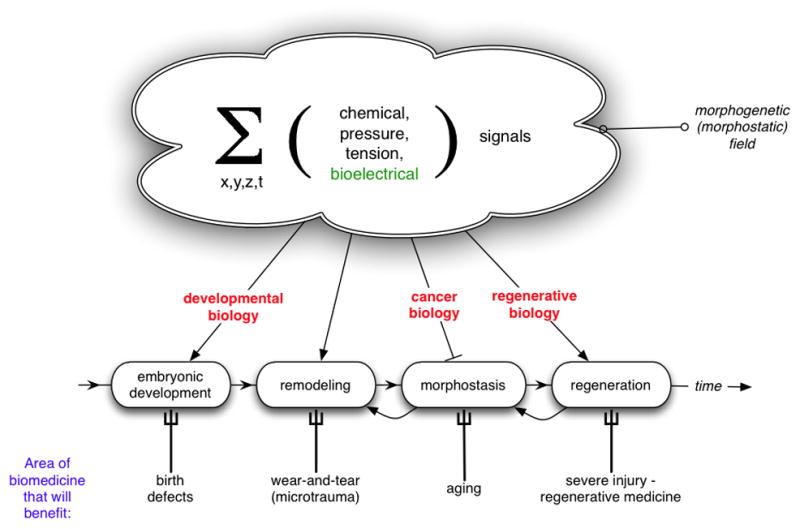
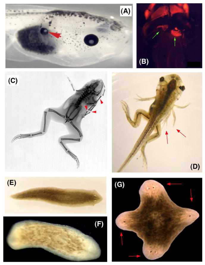
Establishment of shape during embryonic development, and the maintenance of shape against injury or tumorigenesis, requires constant coordination of cell behaviors toward the patterning needs of the host organism. Molecular cell biology and genetics have made great strides in understanding the mechanisms that regulate cell function. However, generalized rational control of shape is still largely beyond our current capabilities. Significant instructive signals function at long range to provide positional information and other cues to regulate organism-wide systems properties like anatomical polarity and size control. Is complex morphogenesis best understood as the emergent property of local cell interactions, or as the outcome of a computational process that is guided by a physically encoded map or template of the final goal state? Here I review recent data and molecular mechanisms relevant to morphogenetic fields: large-scale systems of physical properties that have been proposed to store patterning information during embryogenesis, regenerative repair, and cancer suppression that ultimately controls anatomy. Placing special emphasis on the role of endogenous bioelectric signals as an important component of the morphogenetic field, I speculate on novel approaches for the computational modeling and control of these fields with applications to synthetic biology, regenerative medicine, and evolutionary developmental biology.


Figures

Figure 1
Figure 1. The morphogenetic field in development, regeneration, and neoplasm and its applications to medicine
The morphogenetic field can be defined as the sum, integrated over 1 temporal and 3 spatial dimensions, of all non-local patterning signals impinging on cells and cell groups in an organism. Functionally, long-range signals (such as planar polarity of proteins on cell surfaces, standing waves of gene expression, voltage potential, and tensile forces, and chemical morphogen gradients) carry information about both the existing and the future pattern of the organism. This allows the initial development of complex form from a single fertilized egg cell, as well as the subsequent maintenance of form in adulthood against trauma and individual cell loss. Errors in various aspects of the establishment and interpretation of these fields result in failures to maintain systems-level properties of anatomical shape, manifesting as birth defects, cancer, aging, and failure to regenerate after injury. Thus, almost every area of biomedicine is impacted by our knowledge of how cells interact with and within this set of complex signals.
Figure 2
Figure 2. Voltage gradients in vivo
A: Fluorescent voltage reporter dyes allow characterization of physiological gradients in vivo, such as this image of a 16-cell frog embryo that simultaneously reveals cells’ transmembrane potential levels (blue = hyperpolarized, red = depolarized) in vivo, as well as domains of distinct Vmem around a single blastomere’s surface (compare the side indicated by the yellow arrowhead with the one indicated by the red arrowhead). Provided courtesy of Dany Adams. B: Isopotential cell fields can also demarcate subtle prepatterns existing in tissues, such as the hyperpolarized domains (red arrowheads) that presage the expression of regulatory genes such as Frizzled during frog embryo craniofacial development; these patterns of transmembrane potential are not merely readouts of cell state but are functional determinants of gene expression and anatomy (Vandenberg et al., 2011).
Figure 3
Figure 3. Emergence of complex morphology from simple low-level rules
A: A short function can be defined over a complex variable Z; this function is iterated – applied repeatedly to each result of the previous iteration (Levin, 1994; Mojica et al., 2009; Pickover, 1986). B: A Julia set pattern can be created by iterating such a function for each point in the plane: Z0=X+Yi for coordinates of each point (X,Y). Each point is then assigned a color based on how fast the absolute value of Z exceeds a threshold upon iterated application of the function to the initial Z. This extremely simple algorithm gives rise to a complex morphology, illustrating how spatial complexity can emerge from a simple set of low-level rules without being directly specified or encoded anywhere in those rules. C: While it is easy to produce an image corresponding to a set of rules (A -> B), the inverse problem is much harder. In general, it is impossible to know how to modify the generative rules (A) to give rise to a desired pattern – for example, a modified version of B where one element is rotated 90° (yellow arrow).
Figure 4
Figure 4. Alteration of target morphology
(A) In the red deer Cervus elaphus, an experimental incision in one location induces a slight hypertrophy in the first year but results in a supernumerary (ectopic) tine at that same location in the next year. Image modified (with permission) after Fig. 22 of (Bubenik, 1966). In planaria, the anterior-posterior polarity (head vs. tail) during fragment regeneration can be perturbed by manipulating the flow of ions among cells. When communication is reduced for 48 hours, a fragment will regenerate into a 2-headed form in 7 days (B). Cuts made over months following this treatment, in plain water (no exposure to any perturbation) result in the regeneration of 2-headed worms (C), demonstrating that information present in a dynamic physiological network can be canalized or remembered so that the shape to which the animal regenerates in further rounds of damage (target morphology) is directly and (permanently?) altered without modification of DNA sequence. Planarian images in B courtesy of Junji Morokuma.
Figure 5
Figure 5. Modular alteration of pattern by biophysical modulation
Changing the pattern encoded in physiological networks results in coherent, modular alterations of form in vivo. Gradients of resting transmembrane potential were artificially altered by misexpressing mRNA encoding specific ion channels (in frog embryos) or by pharmacologically manipulating native ion translocator proteins (in planaria). The results in Xenopus laevis embryos include induction of: whole ectopic eyes on the gut (A, red arrow), a complete beating ectopic heart (B, green arrow), and well-formed ectopic limbs with normal bone structure (C,D, red arrows). In regenerating planarian flatworms (normal morphology in E), such modulation can be used to control the anatomical polarity and overall body-plan, including no-head worms (F) and 4-headed worms (G, red arrowheads indicate the heads), all of which are viable.

References

    1. Adamatzky A. Game of Life cellular automata. Springer; London: 2010. p. xix.p. 579.
    1. Adams DS, Levin M. Endogenous voltage gradients as mediators of cell-cell communication: strategies for investigating bioelectrical signals during pattern formation. Cell Tissue Res. 2012a in press. - PMC - PubMed
    1. Adams DS, Levin M. General Principles For Measuring Resting Membrane Potential And Ion Concentration Using Fluorescent Bioelectricity Reporters. Cold Spring Harbor Protocols. 2012b in press. - PMC - PubMed
    1. Adams DS, Levin M. Measuring resting membrane potential using the fluorescent voltage reporters DiBAC4(3) and CC2-DMPE. Cold Spring Harbor Protocols. 2012c in press. - PMC - PubMed
    1. Adams DS, Masi A, Levin M. H+ pump-dependent changes in membrane voltage are an early mechanism necessary and sufficient to induce Xenopus tail regeneration. Development. 2007;134:1323–1335. - PubMed

Publication types