Review
. 2012 Aug;33(4):295-317.
doi: 10.1016/j.mam.2012.04.005. Epub 2012 Apr 21.

Understanding age-related macular degeneration (AMD): relationships between the photoreceptor/retinal pigment epithelium/Bruch's membrane/choriocapillaris complex

Affiliations
Review

Understanding age-related macular degeneration (AMD): relationships between the photoreceptor/retinal pigment epithelium/Bruch's membrane/choriocapillaris complex

Imran Bhutto et al. Mol Aspects Med. 2012 Aug.

Abstract

There is a mutualistic symbiotic relationship between the components of the photoreceptor/retinal pigment epithelium (RPE)/Bruch's membrane (BrMb)/choriocapillaris (CC) complex that is lost in AMD. Which component in the photoreceptor/RPE/BrMb/CC complex is affected first appears to depend on the type of AMD. In atrophic AMD (~85-90% of cases), it appears that large confluent drusen formation and hyperpigmentation (presumably dysfunction in RPE) are the initial insult and the resorption of these drusen and loss of RPE (hypopigmentation) can be predictive for progression of geographic atrophy (GA). The death and dysfunction of photoreceptors and CC appear to be secondary events to loss in RPE. In neovascular AMD (~10-15% of cases), the loss of choroidal vasculature may be the initial insult to the complex. Loss of CC with an intact RPE monolayer in wet AMD has been observed. This may be due to reduction in blood supply because of large vessel stenosis. Furthermore, the environment of the CC, basement membrane and intercapillary septa, is a proinflammatory milieu with accumulation of complement components as well as proinflammatory molecules like CRP during AMD. In this toxic milieu, CC die or become dysfunction making adjacent RPE hypoxic. These hypoxic cells then produce angiogenic substances like VEGF that stimulate growth of new vessels from CC, resulting in choroidal neovascularization (CNV). The loss of CC might also be a stimulus for drusen formation since the disposal system for retinal debris and exocytosed material from RPE would be limited. Ultimately, the photoreceptors die of lack of nutrients, leakage of serum components from the neovascularization, and scar formation. Therefore, the mutualistic symbiotic relationship within the photoreceptor/RPE/BrMb/CC complex is lost in both forms of AMD. Loss of this functionally integrated relationship results in death and dysfunction of all of the components in the complex.


Figures

Figure 1
Figure 1
A cross section of the fovea from a Macaque monkey demonstrates the layers of retina and the morphological relationship of photoreceptor/RPE/BrMb/choroid complex. To the left and right of the foveal pit, the center of macula, the layers of the sensory retina are clearly visible. The inner most neuronal nuclei are of ganglion cells (GCL). The inner plexiform layer (IPL) separates the inner nuclear layer of neurons (INL) from the ganglion cell soma. The outer plexiform layer (OPL) represents the synapses between photoreceptors in the outer nuclear layer (ONL) and secondary neurons in the INL. The photoreceptor inner segments (IS) are mitochondria-rich and their outer segments (OS) make close contact with the retinal pigment epithelium (RPE), the outer most layer of retina. Bruch’s membrane (not discernible at this magnification) separates the RPE from the choriocapillaris (CC). The melanocytes of choroid are the extremely dark structures below the CC.
Figure 2
Figure 2
Alkaline phosphatase (APase)-incubated choroid from a 88 year-old Caucasian male with GA showing the nonatrophic region (A,D&G), border region (B,E&H) and atrophic region (C,F&I) in flat view prior to embedment (A-C) and in cross sections stained with PAS and hematoxylin (D–I). Blue APase reaction product is present in viable blood vessels only and is most prominent in neovascularization. (A) APase stained capillaries in the nonatrophic region have broad diameter lumens filled with serum APase (arrow in D&G), with endothelial cells and pericytes underlying viable RPE (arrowhead in D&G). At the border region, RPE appear hypertrophic (arrowhead in E&H), capillaries appear constricted (arrow in E&H), and some have completely degenerated (paired arrows H). A thin basal laminar deposit is associated with Bruch’s membrane (open arrow). In the atrophic region, many capillaries have degenerated leaving only remnants of basement membrane material (arrows in F&I). (scale bar = 100 μm in A–C, 30 μm in D–F, 10 μm in G–I) [Figure 10 from McLeod et al, Invest. Ophthalmol. Vis. Sci 50:4982-4991, 2009 (McLeod et al., 2009) with permission]
Figure 3
Figure 3
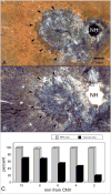
APase-incubated choroid from an 81 year-old Caucasian female with wet AMD. A submacular sea fan-like CNV formation is shown (arrowheads) using epi-illumination to analyze RPE (A) and transillumination (B) to analyze viable blood vessels. Areas of choriocapillaris dropout are located in advance of the CNV (asterisks). Percent RPE and vascular area measurements made in 2 mm intervals from the CNV (C) show that capillary dropout is severe (<20% vascular area) immediately in advance of the sea fan and present well beyond the area with neovascularization. (NH = optic nerve head, scale bar = 2 mm) [Figure 6 from McLeod et al, Invest. Ophthalmol. Vis. Sci 50:4982-4991, 2009 (McLeod et al., 2009) with permission]
Figure 4
Figure 4
APase-incubated choroid from the 81 year-old Caucasian female (higher magnification than shown in Figure 3) with wet AMD showing submacular CNV using epi-illumination (A&C) and transillumination (B&D). The front of growing vessels is closely associated with viable RPE (arrows in A-D). Areas of CC dropout are evident in advance of the CNV (asterisks A&B). In PAS and hematoxylin stained sections, the equatorial region (E&H) has broad capillaries (arrows) containing serum APase with both endothelial cells and pericytes. The RPE has a normal morphology (arrowhead) and Bruch’s membrane is free of deposits. In sections taken 1 mm beyond the CNV (F&I), only a few capillaries are viable (arrows) and many degenerative capillaries are seen (asterisks in I). The RPE is hypertrophic (arrowheads) and a basal laminar deposit is present. Sections taken through the edge of the CNV (G&J) show degenerative capillaries (asterisk in J), sub-RPE neovascularization (open arrow) and hypertrophic RPE overlying the leading edge of the CNV. (scale bar=0.5mm A&B, 100 μm C&D, 30 μm E–G, and 10 μm H–J)[Figure 11 from McLeod et al, Invest. Ophthalmol. Vis. Sci 50:4982-4991, 2009 (McLeod et al., 2009) with permission]
Figure 5
Figure 5
Schematic of a normal RPE/BrMb/CC complex (A) and the changes that occur in dry (B) and wet (C) AMD. (A) The CC (purple) lie under BrMb (Black line) and large choroidal blood vessels (blue/red) are below the CC. RPE reside on top of BrMb. In GA (B), RPE are lost and then the CC become attenuated but some CC survive but are constricted. In wet AMD (C), CC is lost while RPE remain. The RPE become hypoxic and produce hypoxia-inducible growth factors like VEGF, which stimulate the formation of CNV (solid purple blood vessel).
Figure 6
Figure 6
Serial sections of submacular choroid from normal aged control (left) and retina and submacular choroid from an AMD subject (right) incubated with TSP-1, endostatin, and PEDF antibodies. Right panels are high magnification photos of left panels. (A–D) Hematoxylin and eosin (H&E) staining show morphological features of retina and choroid like migration of RPE cells into retina in AMD (C,D). Pigment in immunostained sections was bleached from RPE and choroidal melanocytes. Immunostaining of CD-34 (E–H) is associated with the retinal and choroidal blood vessels including CC (arrow). In aged control choroid, TSP-1 immunoreactivity (I and J) is intense especially in BrMb (arrowhead). Both endostatin (M and N) and PEDF (Q and R) are prominent in RPE basal lamina, BrMb, and CC basement membrane and show similar pattern and intensity of immunostaining. In contrast, expression of TSP-1 (K and L), endostatin (O and P), and PEDF (S and T) is greatly reduced in AMD choroid compared to the aged control and the reaction product of endostatin and PEDF appears more diffuse in choroidal stroma. (arrowhead, Bruch’s membrane; arrow, choriocapillaris).
Figure 7
Figure 7
Graphic representation of data from 8 aged control subjects and 12 AMD subjects, represented by the immunohistochemistry in Figure 6. The scores (0–7) on the Y-axis represent the mean scores from three masked observers. Aged control subject data is represented by open bars and AMD subjects by black bars. A significant difference (***, P<0/01) was found for all three inhibitors in BrMb between control and AMD subjects.
Figure 8
Figure 8
Immunolocalization of C-reactive protein (CRP) and complement factor H (CFH) in submacular choroid from aged control, early and late wet AMD eyes. Periodic acid-Schiff’s (PAS) and hematoxylin (Hem) staining shows morphological features of the choroid from aged control (A), drusen (asterisk) in early AMD (B) and CNV (large arrow) anterior to RPE in wet AMD (C). Pigment in immunostained sections was bleached from RPE and choroidal melanocytes. Immunostaining of CD34 is associated with CC (small arrow) and large choroidal vessels appear morphologically normal with broad lumens in aged control (D), whereas CC lumens appear irregular and constricted in early (E) and wet AMD (F). In aged control choroid, CRP (G) and CFH (J) are prominently localized to the CC, intercapillary septa (ICS) and BrM (open arrowhead). CRP immunoreactivity is significantly increased in early (H) and late AMD (I) choroids compared to the aged control and appear more diffuse in choroidal stroma. CFH in early AMD (K) is comparable to aged control, whereas it is significantly decreased in wet AMD (L). Drusen are intensely labeled with CRP and CFH (H and K). Note that in wet AMD the CNV (large arrow), intensely labeled with CD34 antibody (F), has more CRP and less CFH (I and L). Nonimmune rabbit IgG (NIIgG) yields a very weak to negative reaction product except in drusen (M, N, O). [Figure 3 from Bhutto et al British Journal of Ophthalmology 95:1323-1330, 2011 (Bhutto et al., 2011) with permission]

References

    1. Adamis AP, Shima DT, Yeo KT, Yeo TK, Brown LF, Berse B, D’Amore PA, Folkman J. Synthesis and secretion of vascular permeability factor/vascular endothelial growth factor by human retinal pigment epithelial cells. Biochem Biophys Res Commun. 1993;193 (2):631–638. - PubMed
    1. Age-related Eye Disease Study Research Group. A randomized, placebo-controlled, clinical trial of high-dose supplementation with vitamins C and E, beta carotene, and zinc for age-related macular degeneration and vision loss: AREDS report no. 8. Arch Ophthalmol. 2001;119 (10):1417–1436. - PMC - PubMed
    1. Ahuja P, Caffe AR, Holmqvist I, Soderpalm AK, Singh DP, Shinohara T, van Veen T. Lens epithelium-derived growth factor (LEDGF) delays photoreceptor degeneration in explants of rd/rd mouse retina. Neuroreport. 2001;12 (13):2951–2955. - PubMed
    1. Aiello LP, Avery RL, Arrigg PG, Keyt BA, Jampel H, Shah ST, Pasquale LR, Thieme H, Iwamoto MA, Park JE, Nguyen HV, Aiello LM, Ferrara N, King G. Vascular endothelial growth factor in ocular fluid of patients with diabetic retinopathy and other retinal disorders. N Engl J Med. 1994;331:1480–1487. - PubMed
    1. Aisenbrey S, Zhang M, Bacher D, Yee J, Brunken WJ, Hunter DD. Retinal pigment epithelial cells synthesize laminins, including laminin 5, and adhere to them through alpha3- and alpha6-containing integrins. Invest Ophthalmol Vis Sci. 2006;47 (12):5537–5544. - PMC - PubMed

Publication types