. 2012 May 15;22(5):1079-91.
doi: 10.1016/j.devcel.2012.02.003. Epub 2012 Apr 26.

LTB4 is a signal-relay molecule during neutrophil chemotaxis

Affiliations

LTB4 is a signal-relay molecule during neutrophil chemotaxis

Philippe V Afonso et al. Dev Cell. .

Abstract

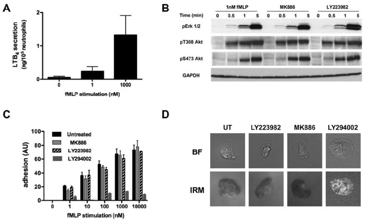
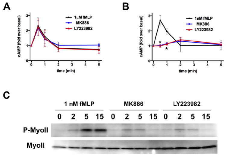
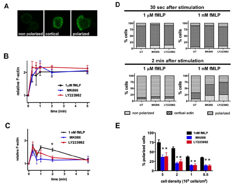
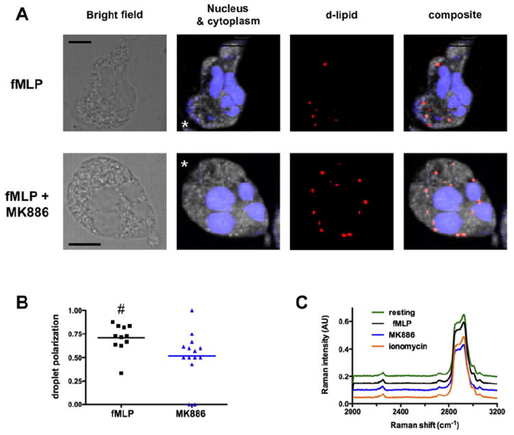
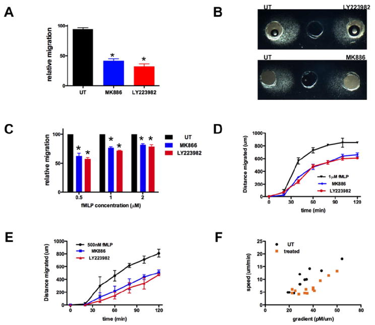
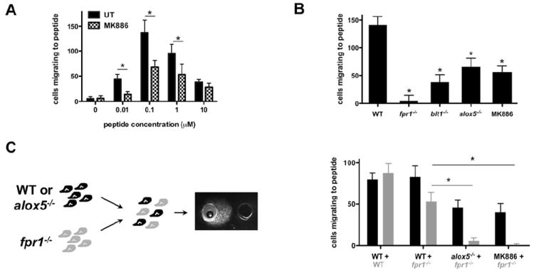
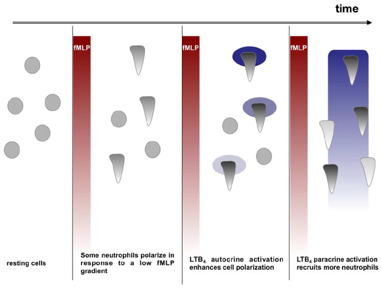
Neutrophil recruitment to inflammation sites purportedly depends on sequential waves of chemoattractants. Current models propose that leukotriene B(4) (LTB(4)), a secondary chemoattractant secreted by neutrophils in response to primary chemoattractants such as formyl peptides, is important in initiating the inflammation process. In this study we demonstrate that LTB(4) plays a central role in neutrophil activation and migration to formyl peptides. We show that LTB(4) production dramatically amplifies formyl peptide-mediated neutrophil polarization and chemotaxis by regulating specific signaling pathways acting upstream of actin polymerization and MyoII phosphorylation. Importantly, by analyzing the migration of neutrophils isolated from wild-type mice and mice lacking the formyl peptide receptor 1, we demonstrate that LTB(4) acts as a signal to relay information from cell to cell over long distances. Together, our findings imply that LTB(4) is a signal-relay molecule that exquisitely regulates neutrophil chemotaxis to formyl peptides, which are produced at the core of inflammation sites.


Figures

Figure 1
Figure 1. LTB4 secretion does not alter fMLP-induced ERK and PI3K activation
A. fMLP-induced LTB4 secretion by neutrophils is dose dependent. Primary human neutrophils were stimulated with fMLP for 1 min, and the amount of LTB4 in the supernatant was determined by ELISA. Results represent the average ± SEM of four independent experiments. B. LTB4 secretion does not amplify Erk1/2 and Akt phosphorylation upon stimulation with sub-saturating doses of fMLP. Primary human neutrophils were stimulated with 1 nM fMLP after pretreatment with either 100 nM MK866, 10 μM LY223982, or DMSO as a control. The western blot for the kinetics of activation is representative of three independent western blot analyses. Also see Fig. S1A-B. C. fMLP-induced LTB4 secretion has no impact on cell adhesion to fibronectin. Primary human neutrophils were treated with either 100 nM MK866, 10 μM LY223982, or 40 μM LY294002, a PI3K inhibitor. Cells were plated on fibronectin-coated plates for 10 min and uniformly stimulated with different concentrations of fMLP. The plates were then shaken and the number of remaining cells attached to the plates was estimated by crystal violet staining. Results represent the average ± SEM of four independent experiments. D. Neutrophil adhesion pattern is not altered upon treatment with LTB4 pathway inhibitors. Neutrophil adhesion to fibronectin-coated plates upon stimulation with 1 nM fMLP was observed by IRM in the presence or absence of drugs as described in panel C. The areas of close contact of neutrophils to the substratum appear dark in the IRM image. Representative images are presented.
Figure 2
Figure 2. LTB4 secretion enhances fMLP-induced cAMP production and MyoII phosphorylation
A. LTB4 secretion has no impact on intracellular cAMP accumulation in neutrophils stimulated with a saturation dose of fMLP. Primary human neutrophils were treated with 100 nM MK886 or 10 μM LY223982 or DMSO as control and stimulated with 1μM fMLP. Intracellular cAMP levels were determined by ELISA at the indicated time points. Results represent the average ± SEM of three independent experiments. B. LTB4 inhibition reduces cAMP accumulation in neutrophils treated with a sub-saturating dose of fMLP. Primary human neutrophils were treated as in panel A and stimulated with 1 nM fMLP. Results represent the average ± SEM of three independent experiments. * p<0.05, ANOVA; Dunnett posthoc test. Also see Fig. S2A-B. C. LTB4 secretion amplifies phosphorylated MyoII levels in response to sub-saturating doses of fMLP. Primary human neutrophils were plated on fibronectin-coated plates for 10 min and stimulated uniformly with 1 nM fMLP in the presence or absence of drugs as described in panel A. The western blot for the kinetics of activation is representative of three independent western blot analyses. Also see Fig. S2C.
Figure 3
Figure 3. Autocrine and paracrine LTB4 secretion enhances fMLP-induced cell polarization
A. Different stages of neutrophil polarization can be observed in response to fMLP stimulation. Primary human neutrophils were plated on gelatin-coated plates. Cells were stimulated, fixed and F-actin was stained with FITC-phalloidin. Representative images are presented. B. LTB4 secretion has no impact on neutrophil response to a saturating dose of fMLP. Primary human neutrophils were treated with 100 nM MK886 or 10 μM LY223982 or DMSO as control, stimulated with 1 μM fMLP, fixed and the F-actin network stained with FITC-phalloidin. The kinetic of the average fluorescence was determined by FACS analysis. Results represent the average ± SEM of three independent experiments. C. Neutrophil treatment with LTB4 inhibitors reduces neutrophil polarization in response to sub-saturating doses of fMLP. Primary human neutrophils were treated as in panel B, stimulated with 1 nM fMLP and F-actin levels were determined by FACS, after staining with FITC-phalloidin. Results represent the average ± SEM of three independent experiments. * p<0.005, ANOVA; Dunnett posthoc test. D. LTB4 amplifies neutrophil polarization after 2 min of fMLP stimulation. Primary human neutrophils were treated as in panel B, plated on gelatin-coated plates, stimulated with fMLP and fixed at different time points. Cells were stained with F-actin and counted into 3 categories (unpolarized, accumulated cortical F-actin, polarized). Results represent the average of four independent experiments. E. LTB4 amplifies neutrophil polarization in an autocrine and paracrine manner. Primary human neutrophils were treated as in panel B, plated on gelatin-coated plates at different cell densities for 10 min. After 2 min stimulation with 1 nM fMLP, cells were fixed and the number of polarized cells was counted. Results represent the average ± SEM of three independent experiments. *p<0.05, ANOVA; Dunnett posthoc test.
Figure 4
Figure 4. Arachidonic acid accumulates at the front of polarized neutrophils
A. Bright field and CARS images of deuterated punctates localized in polarized primary human neutrophils migrating to 1μM fMLP in underagarose assay. Representative images of polarized cells untreated (upper panel) or treated with MK886 (lower panel) are presented. The false-colored chemical images for nucleus (blue), cytoplasm (grey), and deuterated punctates (red) were constructed from Raman intensities at 2952 cm-1 and 2850 cm-1 for nucleus, and intensities at 2900 cm-1 for cytoplasm and 2250 cm-1 for deuterated punctates, respectively. B. Location parameters of deuterated punctates in neutrophils are plotted for two differently treated neutrophils. The location parameter is defined as (number of punctates at the front)/(total number of punctates). Neighboring image pixels (> four pixels) are counted as one regardless of the overall size. # p-value = 0.009, Wilcoxon test. C. Comparison of the CARS spectra of deuterated punctates found in polarized neutrophils under the indicated conditions.
Figure 5
Figure 5. Neutrophil migration to fMLP is amplified by fMLP-induced LTB4 paracrine/autocrine secretion
A. LTB4 secretion amplifies neutrophil migration to fMLP. The number of primary human neutrophils migrating to 1 μM fMLP in a 4 μm transwell was determined after 2 h. Results represent the relative percentage of migrating cells after treatment (average ± SEM) of three independent experiments. *p<0.05, Friedman test; Dunns posthoc test. B. LTB4 secretion amplifies neutrophil chemotaxis to fMLP. Representative images of primary human neutrophils migrating to 1 μM fMLP in the under-agarose assay are shown. C. LTB4 secretion amplifies neutrophil chemotaxis to fMLP. The distance migrated by primary human neutrophils treated with LTB4 pathway inhibitors is compared to the one migrated by untreated cells. Results represent the relative distance migrated (average ± SEM, n=3) in under-agarose assay in 2 h. *p<0.05, Friedman test; Dunns posthoc test. D-E. Kinetics of neutrophil migration in under-agarose assays. The distance migrated by primary human neutrophils to either 1 μM fMLP (D) or 500 nM fMLP (E) was determined at different times points. Results represent the average ± SEM of three independent experiments. F. Impact of LTB4 secretion on neutrophil migration to different fMLP gradients. For each segment of 20 min of migration, the average speed was determined and the local gradient of the front of migration was determined using theoretical charts (see Fig. S4). The resulting different data points (speed vs. gradient) are plotted.
Figure 6
Figure 6. LTB4 is a signal relay molecule for neutrophils
A. MK886 treatment regulates murine neutrophil migration to MKYMVm. Neutrophils isolated from the bone marrow of WT mouse were allowed to migrate to MKYMVm in the under-agarose assay. The number of neutrophils migrating to fMLP was determined after 5 h of migration. Results represent the average ± SEM number of migrating mouse neutrophils in three independent experiments. *p<0.05, Mann-Whitney tests. B. LTB4 secretion amplifies murine neutrophil migration to MKYMVm. Neutrophils isolated from the bone marrow of mice were allowed to migrate to 100 nM MKYMVm in the under-agarose assay. The number of neutrophils migrating was determined after 5 h of migration. Results represent the average ± SEM number of migrating mouse neutrophils in three independent experiments. *p < 0.05, Friedman test; Dunns posthoc test. C. Neutrophils that do not sense MKYMVm can still migrate to MKYMVm when mixed with WT neutrophils secreting LTB4. Neutrophils isolated from fpr1-/- mice were fluorescently labeled and mixed with WT neutrophils (pretreated or not with 100nM MK886) or neutrophils isolated from alox5-/- mice. The number of fluorescent and non-fluorescent cells that migrate to 100 nM MKYMVm was determined after 5 h migration. Results represent the average ± SEM number of migrating mouse neutrophils in three independent experiments. *p < 0.05, Friedman test; Dunns posthoc test.
Figure 7
Figure 7. Model for LTB4 as a signal relay molecule for neutrophils migrating to fMLP
In response to an external fMLP gradient, some neutrophils respond, polarize and release LTB4. The local LTB4 gradient strengthens and stabilizes cell polarization of these first responders. As LTB4 production is fMLP-concentration dependent, a secondary LTB4 gradient is formed parallel to the fMLP gradient. Neutrophils that were not initially responsive to fMLP, sense the secondary gradient of LTB4 and migrate up this gradient towards the fMLP source, thus amplifying the inflammatory response.

References

    1. Alessi DR, James SR, Downes CP, Holmes AB, Gaffney PR, Reese CB, Cohen P. Characterization of a 3-phosphoinositide-dependent protein kinase which phosphorylates and activates protein kinase Balpha. Curr Biol. 1997;7:261–269. - PubMed
    1. Berger M, Budhu S, Lu E, Li Y, Loike D, Silverstein SC, Loike JD. Different G(i)-coupled chemoattractant receptors signal qualitatively different functions in human neutrophils. J Leukoc Biol. 2002;71:798–806. - PubMed
    1. Boxio R, Bossenmeyer-Pourie C, Steinckwich N, Dournon C, Nusse O. Mouse bone marrow contains large numbers of functionally competent neutrophils. J Leukoc Biol. 2004;75:604–611. - PubMed
    1. Brito GA, Souza MH, Melo-Filho AA, Hewlett EL, Lima AA, Flores CA, Ribeiro RA. Role of pertussis toxin A subunit in neutrophil migration and vascular permeability. Infect Immun. 1997;65:1114–1118. - PMC - PubMed
    1. Canetti CA, Leung BP, Culshaw S, McInnes IB, Cunha FQ, Liew FY. IL-18 enhances collagen-induced arthritis by recruiting neutrophils via TNF-alpha and leukotriene B4. J Immunol. 2003;171:1009–1015. - PubMed

Publication types