. 2012 Jun;33(6):817-22.
doi: 10.1038/aps.2012.30. Epub 2012 Apr 30.

E-cadherin promotes proliferation of human ovarian cancer cells in vitro via activating MEK/ERK pathway

Affiliations

E-cadherin promotes proliferation of human ovarian cancer cells in vitro via activating MEK/ERK pathway

Ling-ling Dong et al. Acta Pharmacol Sin. 2012 Jun.

Abstract

Aim: E-cadherin is unusually highly expressed in most ovarian cancers. This study was designed to investigate the roles of E-cadherin in the carcinogenesis and progression of ovarian cancers.

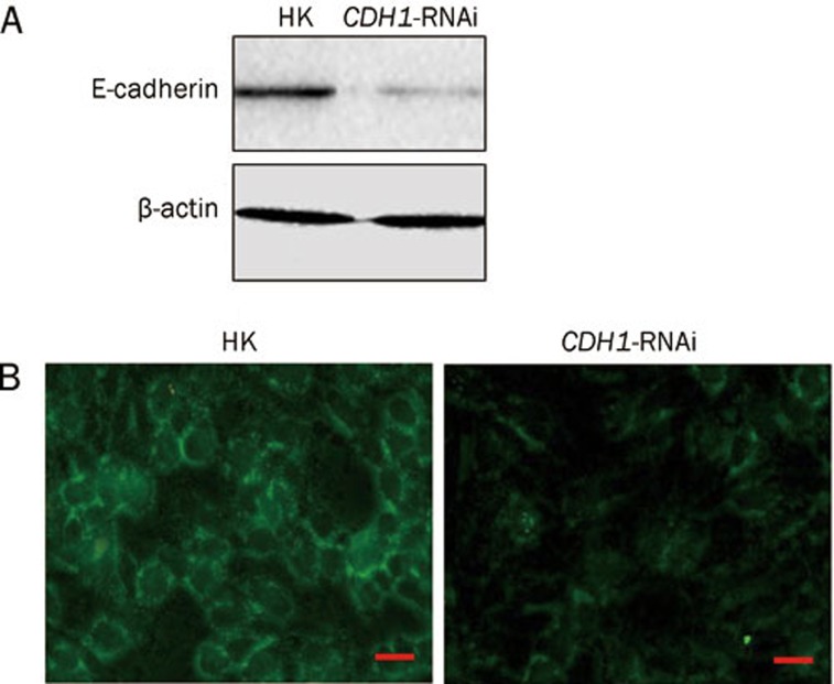
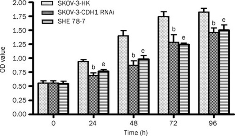
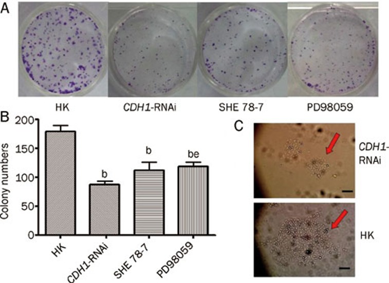
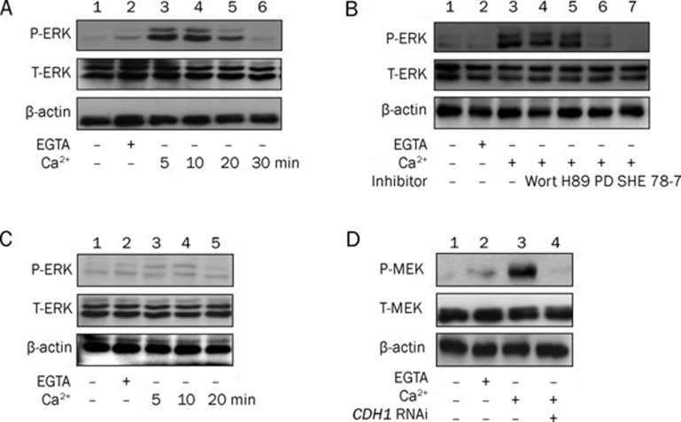
Methods: Human ovarian adenocarcinoma cell line SKOV-3 was examined. E-cadherin gene CDH1 in SKOV-3 cells was knocked down via RNA interference (RNAi), and the resultant variation of biological behavior was observed using CCK-8 and colony formation experiment. E-cadherin-mediated Ca(2+)-dependent cell-cell adhesion was used to study the mechanisms underlying the effects of E-cadherin on the proliferation and survival of SKOV-3 cells. The expression levels of E-cadherin, extracellular signal-related kinase (ERK), phosphorylated ERK (P-ERK) were measured using Western blot assays.

Results: Transfection with CDH1-siRNA for 24-96 h significantly suppressed the growth and proliferation of SKOV-3 cells. E-cadherin-mediated calcium-dependent cell-cell adhesion of SKOV-3 cells resulted in a rapid increase of P-ERK, but did not modify the expression of ERK protein. The phosphorylation of ERK in the cells was blocked by pretreatment with the MEK1 specific inhibitor PD98059 (50 μmol/L), but not by the PI3K inhibitor wortmannin (1 μmol/L) or PKA inhibitor H89 (10 μmol/L).

Conclusion: E-cadherin may function as a tumor proliferation enhancer via activating the MEK/ERK pathway in development of ovarian epithelial cancers.


Figures

Figure 1
Figure 1
RNA interference of CDH1 gene inhibits E-cadherin expression in SKOV-3 cells. (A) Western blot assay shows that E-cadherin protein level in SKOV-3 cells was markedly reduced 48 h after the transfection of CDH1-siRNA plasmids compared with control plasmid HK. (B) Immunofluorescence staining assay using anti-E-cadherin antibody demonstrates the decrease of E-cadherin protein staining on the cell membrane after RNA interference of CDH1 (magnification of ×40). The scale bar stands for 20 μm.
Figure 2
Figure 2
Shutdown of E-cadherin inhibits the proliferation of SKOV-3 cells. After transfection of the CDH1-siRNA and the control plasmids, the proliferation ability of SKOV-3 was monitored by CCK8 assay, at 0, 24, 48, 72 and 96 h, respectively. SKOV-3 cells incubated in the presence of SHE 78-7, a neutral antibody of E-cadherin, were treated in the same way. Assay for every time points was repeated 3 times and the mean±SD of the OD reading value was shown in the figure. The difference of OD values between the control group and CDH1-siRNA group or SHE 78-7 group at every time point was analyzed by student's t-test. bP<0.05 compared with the HK group. eP<0.05 compared with the CDH1-siRNA group.
Figure 3
Figure 3
Inhibition of E-cadherin or MEK suppresses the colony formation ability of SKOV-3 cells. (A, B) Representative result and the quantitative analysis of colony formation assay in SKOV-3 cells showed the obviously decreased colony numbers in not only CDH1-siRNA group but also the SHE 78-7 group and PD98059 group, in contrast to the HK group. Mean±SD of three independent experiments was shown. PD98059, a MEK1 inhibitor. bP<0.05 compared with the HK group, eP<0.05 compared with the CDH1-siRNA group. (C) Colony of SKOV3 cells 48 h after transfection with CHD1-siRNA plasmid displayed the smaller sizes and contained much less cells compared with HK group, the negative control (magnification of ×10). The scale bar stands for 200 μm.
Figure 4
Figure 4
Cell-adhesion-mediated MEK/ERK activation is E-cadherin dependent in SKOV-3 cells. (A) E-cadherin-mediated cell adhesion activates ERK during the time course of calcium restoration in SKOV-3 cells. P-ERK, phosphorylated ERK. T-ERK, total ERK. (B) E-cadherin is required for cell-cell adhesion mediated MEK-ERK activation. Wort, wortmannin (1 μmol/L). H89, PKA inhibitor, 10 μmol/L in concentration. PD, PD98059, MEK1 inhibitor, 50 μmol/L in concentration. SHE 78-7, E-cadherin-specific antibody, 20 μg/mL in concentration. (C, D) After E-cadherin expression was inhibited by siRNA, MEK/ERK failed to be activated after calcium restoration in SKOV-3 cells, for neither ERK (C) nor MEK (D, lane 4) was observed to be phosphorylated. P-MEK, phosphorylated MEK. T-MEK, total MEK.

References

    1. Fang K, Mukhopadhyay T, Shih SH. Retinoic acid modulates epidermal growth factor receptor expression in human lung epithelial cancer cells. J Biomed Sci. 1995;2:256–62. - PubMed
    1. Kemler R. From cadherins to catenins: cytoplasmic protein interactions and regulation of cell adhesion. Trends Genet. 1993;9:317–21. - PubMed
    1. Takeichi M. Cadherin cell adhesion receptors as a morphogenetic regulator. Science. 1991;251:1451–5. - PubMed
    1. Aberle H, Schwartz H, Kemler R. Cadherin-catenin complex: protein interactions and their implications for cadherin function. J Cell Biochem. 1996;61:514–23. - PubMed
    1. Gottardi CJ, Wong E, Gumbiner BM. E-cadherin suppresses cellular transformation by inhibiting beta-catenin signaling in an adhesion-independent manner. J Cell Biol. 2001;153:1049–60. - PMC - PubMed

Publication types