. 2012 Jun;33(6):743-51.
doi: 10.1038/aps.2012.14. Epub 2012 Apr 30.

The regulation of N-terminal Huntingtin (Htt552) accumulation by Beclin1

Affiliations

The regulation of N-terminal Huntingtin (Htt552) accumulation by Beclin1

Jun-chao Wu et al. Acta Pharmacol Sin. 2012 Jun.

Abstract

Aim: Huntingtin protein (Htt) was a neuropathological hallmark in human Huntington's Disease. The study aimed to investigate whether the macroautophagy regulator, Beclin1, was involved in the degradation of Htt.

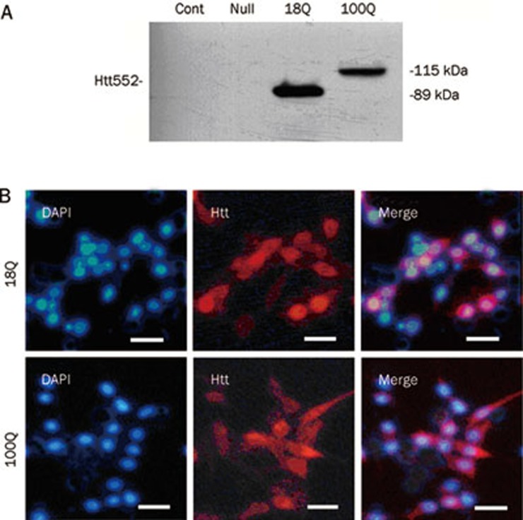
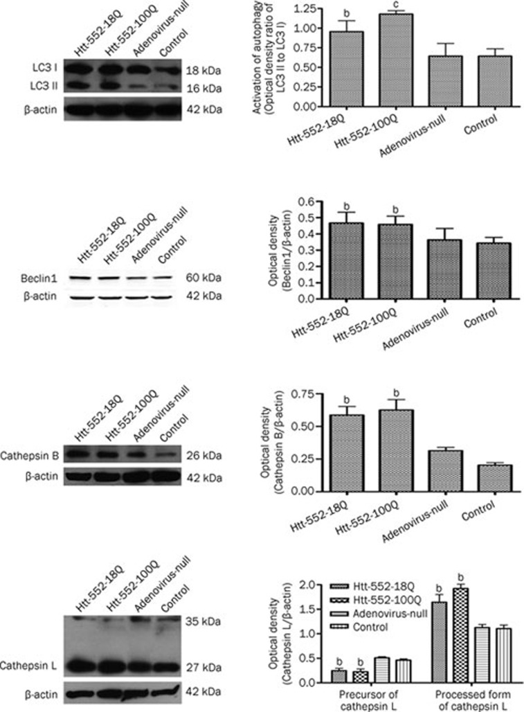
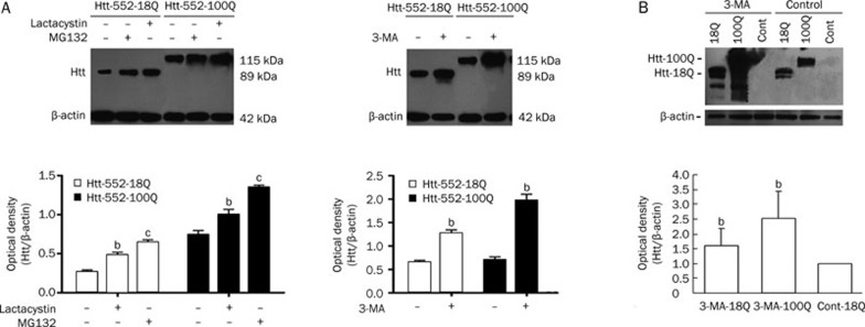
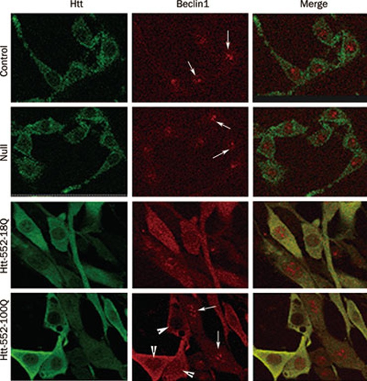
Methods: PC12 cells and primary cultured brain neurons of rats were examined. pDC316 adenovirus shuttle plasmid was used to mediate the expression of wild-type Htt-18Q-552 or mutant Htt-100Q-552 in PC12 cells. The expression of the autophagy-related proteins LC3 II and Beclin1, as well as the lysosome-associated enzymes Cathepsin B and L was evaluated using Western blotting. The locations of Beclin1 and Htt were observed with immunofluorescence and confocal microscope.

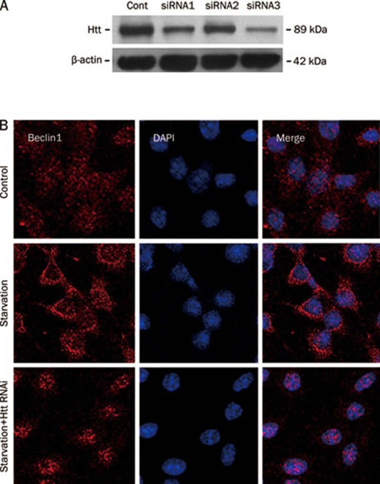
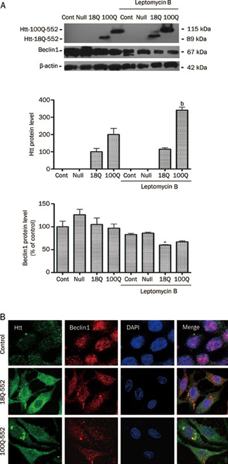
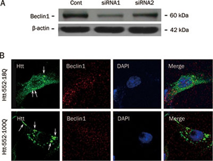
Results: Htt552 expression increased the expression of LC3 II, Beclin1, cathepsin B and L in autophagy/lysosomal degradation pathway. Treatment with the autophagy inhibitor 3-MA or the proteasome inhibitors lactacystin and MG-132 increased Htt552 levels in PC12 cells infected with Ad-Htt-18Q-552 or Ad-Htt-100Q-552. The proteasome inhibitor caused a higher accumulation of Htt552-18Q than Htt552-100Q, and the autophagy inhibitor resulted in a higher accumulation of Htt552-100Q than Htt552-18Q. Similar results were observed in primary cultured neurons infected with adenovirus. In Htt552-expressing cells, Beclin1 was redistributed from the nucleus to the cytoplasm. Htt siRNA prevented Beclin1 redistribution in starvation conditions. Blockade of Beclin1 nuclear export by leptomycin B or Beclin1 deficiency caused by RNA interference induced the formation of mHtt552 aggregates.

Conclusion: Beclin1 regulates the accumulation of Htt via macroautophagy.


Figures

Figure 1
Figure 1
The expression of Htt552 in PC12 cells. PC12 cells were infected with Ad-Htt-18Q-552, Ad-Htt-100Q-552, or Ad-null at MOI 30 and harvested 24 h later for Western blot analysis. Both wild-type and mutant Htt552 were expressed at the expected molecular mass (A). PC12 cells were infected with Ad-Htt-18Q-552, Ad-Htt-100Q-552 at MOI 30 for 24 h and then processed for double immunofluorescence of Htt (red) and the nuclear marker DAPI (blue). Cells were examined with fluorescent microscopy (400×, scale bar=10 μm). Both wild-type and mutant Htt552 had a high expression of Htt552, and the rate of transfection reached approximately 60%–70%.
Figure 2
Figure 2
Autophagy was activated by Htt552 expression. PC12 cells were infected with Ad-Htt-18Q-552, Ad-Htt-100Q-552, or Ad-null at MOI 30 and harvested 24 h later for Western blot analysis. The protein levels of LC3 I and LC3 II increased after Htt552 expression. The data are expressed as a ratio of LC3 II/LC3 I. Beclin1, cathepsin B and the processed form of cathepsin L (bottom panel) were also upregulated after Htt552 expression. The densities of protein bands were analyzed with an image analyzer (Sigma Scan Pro 5) and normalized to the loading control (β-actin). The data are represented as the mean±SEM. Statistical comparisons were carried out with an ANOVA followed by a Dunnett's test. bP<0.05, cP<0.01 vs control.
Figure 3
Figure 3
Both the UPS and ALP play a role in the clearance of Htt552. PC12 cells were infected with Ad-Htt-18Q-552, Ad-Htt-100Q-552, or Ad-null at MOI 30, and primary neurons were infected at MOI 100. After 24 h, cells were treated with the proteasome inhibitors, MG132 and lactacystin, or the autophagy inhibitor, 3-MA, for 18 h. Cells were then lysed for Western blot analysis. Treatment with MG-132, lactacystin (left panel in A) and 3-MA (right panel in A) increased the levels of Htt552. Treatment with 3-MA also increased the levels of Htt552 in rat primary cultured neurons (B). The densities of protein bands were analyzed with an image analyzer (Sigma Scan Pro 5) and normalized to the loading control (β-actin). The data are represented as the mean±SEM. Statistical comparisons were carried out with an ANOVA followed by a Dunnett's test. bP<0.05, cP<0.01 vs control.
Figure 4
Figure 4
Beclin1 levels increased after Htt552 expression in PC12 cells. PC12 cells expressing Htt552 were fixed with pre-cooled EtOH and processed for double immunofluorescence of Htt552 (green) and Beclin1 (red). Cells were analyzed with a confocal microscope (600×). After Htt552 expression, Beclin1 was upregulated in the cytoplasm but reduced in the nucleus, indicating the activation of Beclin1 and the presence of autophagy. The thin arrow refers to Beclin1 in the nucleus, and the arrowhead refers to the export of Beclin1 to the cytoplasm.
Figure 5
Figure 5
The effect of Htt on the activation of Beclin1. PC12 cells were transfected with Htt siRNAs for 6 h. They were then infected with Ad-Htt-18Q-552 at MOI 30 and harvested 24 h later for Western blot analysis. siRNA3 was the most efficient of the samples tested (A). Next, PC12 cells were transfected with Htt siRNA in normal culture medium with 10% FBS for 24 h and then changed to fresh medium without FBS and cultured for another 24 h. Lastly, cells were harvested for immunofluorescence. Knockdown of Htt retained Beclin1 in the nucleus during starvation conditions (B). This result suggests that the activation of Beclin1 was inhibited.
Figure 6
Figure 6
Leptomycin B induced the formation of Htt552 aggregates. PC12 cells were infected with Ad-Htt-18Q-552, Ad-Htt-100Q-552, or Ad-null at MOI 30 and were then treated with leptomycin B for 18 h. Western blot results showed that leptomycin B inhibited the increase of Beclin1 after Htt552 expression (A). The densities of protein bands were analyzed with an image analyzer (Sigma Scan Pro 5) and normalized to the loading control (β-actin). The data are represented as the mean±SEM. Statistical comparisons were carried out using an ANOVA followed by Dunnett's test. bP<0.05 vs cells without leptomycin B treatment. Immunofluorescence results showed that leptomycin B restrained Beclin1 (red) in the nucleus (B). Protein aggregates of mutant Htt552 (green) were observed in some cells. Cells were detected with a confocal microscope (×600).
Figure 7
Figure 7
Beclin1 RNAi induced the formation of Htt552 aggregates. To detect the efficiency of Beclin1 RNAi, PC12 cells were transfected with Beclin1 siRNAs and harvested 24 h later for Western blot analysis. siRNA1 was the most efficient of the samples tested (A). PC12 cells were transfected with Beclin1 siRNA1 for 6 h and were then infected with Ad-Htts. After 48 h, cells were fixed and processed for double immunofluorescence of Htt (green) and the nuclear marker DAPI (blue). Cells were analyzed with a confocal microscope (×600). Mutant Htt552 clearly formed aggregates after Beclin1 levels were reduced (B).

References

    1. Kim YJ, Yi Y, Sapp E, Wang Y, Cuiffo B, Kegel KB, et al. Caspase 3-cleaved N-terminal fragments of wild-type and mutant Htt are present in normal and Huntington's disease brains, associate with membranes, and undergo calpain-dependent proteolysis. Proc Natl Acad Sci USA. 2001;98:12784–9. - PMC - PubMed
    1. Qin ZH, Gu ZL, Lin F. The advancement of molecular pathology of Huntington's disease. Chin Pham Bull. 2004;20:378–82.
    1. Gauthier LR, Charrin BC, Borrell-Pages M, Dompierre JP, Rangone H, Cordelieres FP, et al. Huntingtin controls neurotrophic support and survival of neurons by enhancing BDNF vesicular transport along microtubules. Cell. 2004;118:127–38. - PubMed
    1. Rigamonti D, Bauer JH, De-Fraja C, Conti L, Sipione S, Sciorati C, et al. Wild-type huntingtin protects from apoptosis upstream of caspase-3. J Neurosci. 2000;20:3705–13. - PMC - PubMed
    1. Nasir J, Floresco SB, O'Kusky JR, Diewert VM, Richman JM, Zeisler J, et al. Targeted disruption of the Huntington's disease gene results in embryonic lethality and behavioral and morphological changes in heterozygotes. Cell. 1995;81:811–23. - PubMed

Publication types

MeSH terms