Review
. 2012 Sep;8(3):629-57.
doi: 10.1007/s11302-012-9300-0. Epub 2012 May 1.

Pathophysiology of astroglial purinergic signalling

Affiliations
Review

Pathophysiology of astroglial purinergic signalling

Heike Franke et al. Purinergic Signal. 2012 Sep.

Abstract

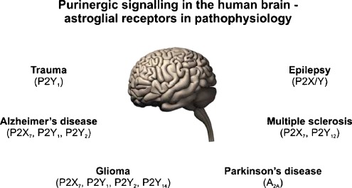
Astrocytes are fundamental for central nervous system (CNS) physiology and are the fulcrum of neurological diseases. Astroglial cells control development of the nervous system, regulate synaptogenesis, maturation, maintenance and plasticity of synapses and are central for nervous system homeostasis. Astroglial reactions determine progression and outcome of many neuropathologies and are critical for regeneration and remodelling of neural circuits following trauma, stroke, ischaemia or neurodegenerative disorders. They secrete multiple neurotransmitters and neurohormones to communicate with neurones, microglia and the vascular walls of capillaries. Signalling through release of ATP is the most widespread mean of communication between astrocytes and other types of neural cells. ATP serves as a fast excitatory neurotransmitter and has pronounced long-term (trophic) roles in cell proliferation, growth, and development. During pathology, ATP is released from damaged cells and acts both as a cytotoxic factor and a proinflammatory mediator, being a universal "danger" signal. In this review, we summarise contemporary knowledge on the role of purinergic receptors (P2Rs) in a variety of diseases in relation to changes of astrocytic functions and nucleotide signalling. We have focussed on the role of the ionotropic P2X and metabotropic P2YRs working alone or in concert to modify the release of neurotransmitters, to activate signalling cascades and to change the expression levels of ion channels and protein kinases. All these effects are of great importance for the initiation, progression and maintenance of astrogliosis-the conserved and ubiquitous glial defensive reaction to CNS pathologies. We highlighted specific aspects of reactive astrogliosis, especially with respect to the involvement of the P2X(7) and P2Y(1)R subtypes. Reactive astrogliosis exerts both beneficial and detrimental effects in a context-specific manner determined by distinct molecular signalling cascades. Understanding the role of purinergic signalling in astrocytes is critical to identifying new therapeutic principles to treat acute and chronic neurological diseases.


Figures

Fig. 1
Fig. 1
From astroglial cell cultures to the human brain—up-regulation of P2Y1R expression on activated GFAP-astrocytes. Confocal images of double immunofluorescence to characterise the expression of P2Y1R subtypes on activated astrocytes after mechanical injury in af in vitro, gi in vivo in rat brain and jl in vivo in human brain. Under physiological conditions (data not shown) in rat and human tissue the expression of P2Y1R on single astrocytes was observed, but with low intensity. In vitro (primary cultures of rat cortical astrocytes): a, d overview about GFAP-positive astrocytes after mechanical injury (marked by asterisks in a; a near the lesion, d far away from the lesion). b, c Single outgrowing astrocytes in the lesioned area with clear co-expression of the astrocytic marker GFAP with the P2Y1R subtype (arrows); d a high number of GFAP/P2Y1R-positive astrocytes at sites remote from the lesion; e, f one example of the co-localisation in higher magnification. In vivo (rat brain, cortex, perilesion area): g overview about the perilesion area after mechanical injury (stab wound injury; according [100]; marked by asterisks) in rat cortex. h, i Single activated GFAP-positive astrocytes in higher magnification with clear co-expression with the P2Y1R subtype (arrows). In vivo (human brain; removed at autopsy, pericontusion zone, cortex): jl After traumatic brain injury (j, overview, pericontusion zone), the P2Y1R was found on activated GFAP-positive astrocytes (k, l; arrows) in the pericontusion zone around the injured area (K. Bremicker, M. Weber and H. Franke, unpublished data)
Fig. 2
Fig. 2
Schematic illustration of examples of signal transduction pathways in astroglial cells following P2X7R activation. After channel opening the P2X7R is permeable for Na+, K+ and Ca2+. Activation of the P2X7R triggers the efflux of K+ from cells and activates IL-1 converting enzyme, leading to cleavage of pro-IL-1β to mature IL-1β and release from the cell. Many events downstream of P2X7R activation are dependent on extracellular calcium influx. Stimulation of ionotropic P2X7Rs leads to activation of phospholipases A2 and D (PLA2, D) and protein kinase C (PKC), e.g. resulting in the activation of glycogen synthase kinase 3 (GSK3) or the activation of caspase cascades. Furthermore, the induction of second messenger and enzyme cascades promoted e.g. the activation of mitogen activated protein kinase (MAPK) pathway proteins (ERK1/2), p38 MAPK, and c-Jun N-terminal kinase (JNK) as well as PI3K/Akt activation. The activity of transcription factors, such as nuclear factor κB (NF-κB), cyclic element-binding protein (CREB), and activator protein (AP-1) are also up-regulated, leading to the expression of proinflammatory genes, such as cyclooxygenase-2 (COX-2) or inducible nitric oxide synthase (iNOS); this in turn causes the production of arachidonic acid (AA) or nitric oxide (NO), respectively. Finally, the release of ATP via pannexin-1 (Panx1) hemichannels as well as of ATP and glutamate via P2X7Rs was also found to take place. The present data suggest that astroglial P2X7R stimulation is associated with neurological disorders leading to neuroinflammation, and apoptosis. The inset summarises examples of P2X7R mediated effects in astrocytes (artwork by courtesy of Dr. Jens Grosche)
Fig. 3
Fig. 3
Schematic illustration of examples of signal transduction pathways in astroglial cells following P2Y1R activation. Stimulation of P2Y1Rs leads to the activation of phospholipases A2 and C (PLA2, C) and protein kinase C (PKC), as well as an increase in intracellular calcium ([Ca2+]i). The activation of P2Y1Rs result in the induction of second messenger and enzyme cascades, e.g. activation of the mitogen activated protein kinase (MAPK) pathway proteins (ERK1/2), p38 MAPK, c-Jun N-terminal kinase (JNK), and PI3K/Akt activation. P2Y1R-mediated signal transducer and activator of transcription 3 (STAT3) signalling may play a role in astrocyte proliferation and reactive astrogliosis. P2Y1R activation appeared to be involved in the activation of caspase (Casp) cascades and the release of arachidonic acid and increase in prostaglandin E2 (PGE2) levels. In addition, P2Y1R activation induces the activity of transcription factors such as nuclear factor κB (NF-κB), cyclic element-binding protein (CREB) and activator protein (AP-1) (which up regulate the expression of proinflammatory genes, e.g. c-Fos, c-Jun, c-Myc). Interaction between adenosine A1 and P2Y1Rs may alter the nucleotide signalling cascades. Modulation of astrocytic P2Y1Rs by the C-terminal domain of the gap junction protein connexin-43 (Cx43) appears to be involved in release of ATP and glutamate. The present data suggest that astroglial P2Y1R stimulation is associated with neurological disorders leading to neuroinflammation, and apoptosis. The inset summarises examples of P2Y1R-mediated effects in astrocytes (artwork by courtesy of Dr. Jens Grosche)
Fig. 4
Fig. 4
Changes of c-Fos expression after stab wound injury in the NAc of rats. a Time course of c-Fos expression 2 h, 4 h, 24 h, 48 h and 4 days after ACSF- and ADPβS-microinjection in comparison to untreated controls (c) and plotted against log time (hours). b Schematic illustration of the localisation of the stab wound in the NAc of rats and the area (square) used for quantification of labelled cells (according to [127]; NAcc, core; NAcs, shell). c Quantification of the effects of ACSF, ADPβS (100 μM), PPADS (30 μM), PD98059 (50 μM; a MAPK inhibitor), wortmannin (Wo, 40 μM; a PI3K inhibitor), and the combination of ADPβS with the respective inhibitors, on the number of c-Fos-positive cells in the NAc of rats after a post-injection time of 2 h. The values are expressed as a percentage of ACSF-treated controls and represent the mean±SEM of five animals per group (*P<0.05, vs. ACSF group; +P<0.05, agonist ADPβS vs. agonist ADPβS/inhibitor group; #P<0.05, inhibitor vs. agonist/inhibitor group). d–g Confocal images of triple-immunofluorescence of d the P2Y1R subtype, e c-Fos-labelling and f GFAP-positivity (g, merge); thick arrow co-localisation with c-Fos; thin arrow no co-localisation with c-Fos. Scale bar: cf = 20 μm
Fig. 5
Fig. 5
Schematic diagram illustrating examples of P2X/YR expression in human brain on glial cells (especially astrocytes) in CNS neuropathological events (artwork by courtesy of Dr. Jens Grosche). For details, see also the Table 3

References

    1. Butt AM, Hamilton N, Hubbard P, et al. Synantocytes: the fifth element. J Anat. 2005;207:695–706. - PMC - PubMed
    1. Kettenmann H, Ransom BR (2005) Neuroglia. Oxford University Press, Oxford, p. 601
    1. Verkhratsky A, Butt A. Glial neurobiology. A textbook. Chichester: Wiley; 2007. p. 220.
    1. Franke H, Illes P. Involvement of P2 receptors in the growth and survival of neurons in the CNS. Pharmacol Ther. 2006;109:297–324. - PubMed
    1. Burnstock G. Physiology and pathophysiology of purinergic neurotransmission. Physiol Rev. 2007;87:659–797. - PubMed

Publication types

MeSH terms

Substances

LinkOut - more resources