

Protein dynamics viewed by hydrogen exchange
- PMID: 22544544
- PMCID: PMC3403437
- DOI: 10.1002/pro.2081
Protein dynamics viewed by hydrogen exchange
Abstract
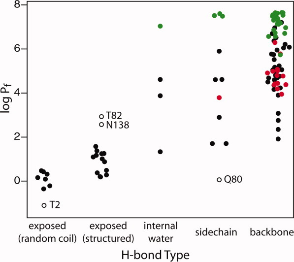
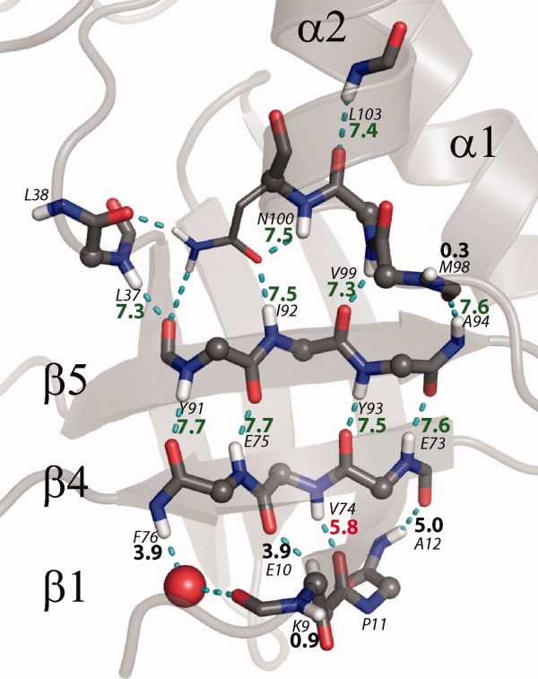
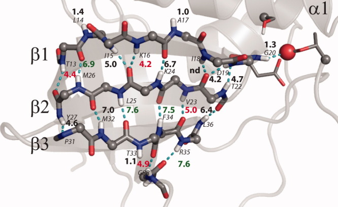
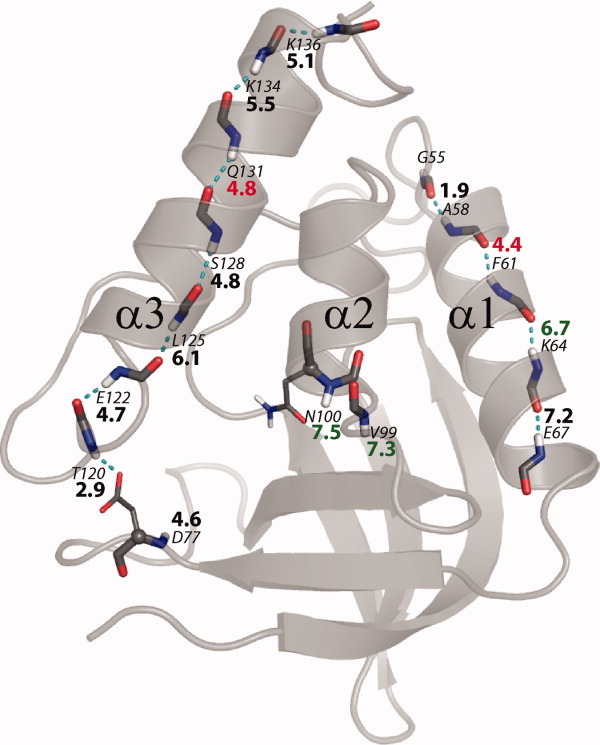
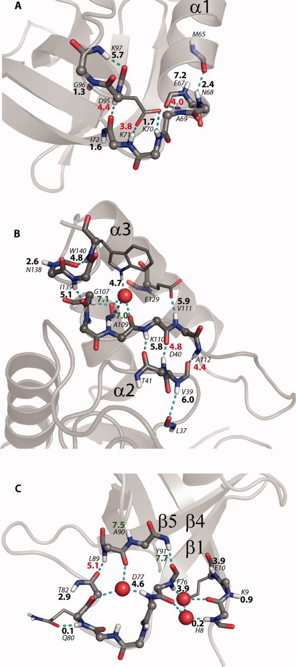
To examine the relationship between protein structural dynamics and measurable hydrogen exchange (HX) data, the detailed exchange behavior of most of the backbone amide hydrogens of Staphylococcal nuclease was compared with that of their neighbors, with their structural environment, and with other information. Results show that H-bonded hydrogens are protected from exchange, with HX rate effectively zero, even when they are directly adjacent to solvent. The transition to exchange competence requires a dynamic structural excursion that removes H-bond protection and allows exposure to solvent HX catalyst. The detailed data often make clear the nature of the dynamic excursion required. These range from whole molecule unfolding, through smaller cooperative unfolding reactions of secondary structural elements, and down to local fluctuations that involve as little as a single peptide group or side chain or water molecule. The particular motion that dominates the exchange of any hydrogen is the one that allows the fastest HX rate. The motion and the rate it produces are determined by surrounding structure and not by nearness to solvent or the strength of the protecting H-bond itself or its acceptor type (main chain, side chain, structurally bound water). Many of these motions occur over time scales that are appropriate for biochemical function.
Copyright © 2012 The Protein Society.
Figures

References
-
- Konermann L, Pan J, Liu YH. Hydrogen exchange mass spectrometry for studying protein structure and dynamics. Chem Soc Rev. 2011;40:1224–1234. - PubMed
-
- Engen J, Jorgensen TJD. Hydrogen exchange mass spectrometry special issue. Int J Mass Spectrom. 2011;302:1–2.
-
- Woodward C, Li RH. The slow-exchange core and protein folding. TIBS. 1998;23:379–379. - PubMed
-
- Truhlar SME, Croy CH, Torpey JW, Koeppe JR, Komives EA. Solvent accessibility of protein surfaces by amide H/H2 exchange MALDI-TOF mass spectrometry. J Am Soc Mass Spectrom. 2006;17:1490–1497. - PubMed
Publication types
MeSH terms
Substances
Grants and funding
LinkOut - more resources
Full Text Sources
