. 2012 Oct;470(10):2827-35.
doi: 10.1007/s11999-012-2370-z. Epub 2012 Apr 28.

Acetabular tilt correlates with acetabular version and coverage in hip dysplasia

Affiliations

Acetabular tilt correlates with acetabular version and coverage in hip dysplasia

Masanori Fujii et al. Clin Orthop Relat Res. 2012 Oct.

Abstract

Background: The rotational position of the acetabulum to the pelvis (acetabular tilt) may influence acetabular version and coverage of the femoral head. To date, the pathologic significance of acetabular tilt in hip dysplasia is unknown.

Questions/purposes: We determined whether acetabular tilt in hip dysplasia is different from that in normal hips and whether this correlates with acetabular version and coverage.

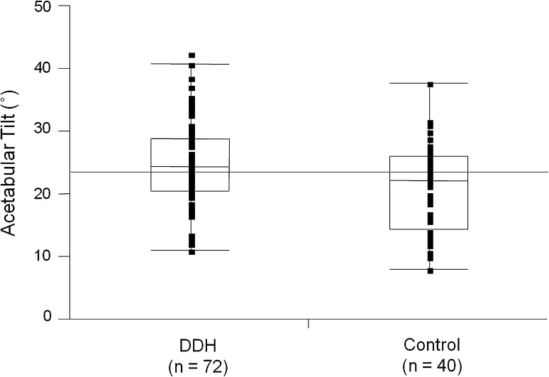
Methods: We measured the acetabular tilt angle on the lateral view of three-dimensional pelvic CT images of 40 patients (72 hips) with hip dysplasia. Forty normal hips from 40 patients were used as controls. The acetabular sector angle was measured as an index for acetabular coverage of the femoral head.

Results: The mean acetabular tilt angle was increased in dysplastic hips compared with controls. In dysplastic hips, a posteriorly rotated acetabulum (increased acetabular tilt) was associated with increased acetabular anteversion and with decreased anterior and anterosuperior acetabular coverage. No correlation was found in controls. In dysplastic hips with a posterior acetabular deficiency, the acetabulum was rotated anteriorly (decreased acetabular tilt) compared with hips with anterior and lateral deficiencies.

Conclusions: We observed a correlation between the rotational position of the acetabulum in the pelvis with acetabular version and coverage in hip dysplasia. Our observations confirmed anterior rotation of the acetabular fragment during periacetabular osteotomies is an anatomically reasonable maneuver for hips with anterolateral acetabular deficiencies, while the maneuver can exacerbate posterior coverage and should be avoided in hips with a posterior acetabular deficiency.

Level of evidence: Level IV, diagnostic study. See the Guidelines for Authors for a complete description of levels of evidence.


Figures

Fig. 1
Fig. 1
A lateral view 3D CT image of the pelvis shows measurement of acetabular tilt angle. The acetabular tilt angle (Angle a) was measured as an angle formed by the intersection of the APP (Line b) passing through the anterior superior iliac spines and pubic tubercles and a line (Line c) connecting the center of an ellipse fitted to the acetabular rim (black dot) and the midpoint of the acetabular notch (white dot).
Fig. 2A–C
Fig. 2A–C
(A) The acetabular anteversion angle (Angle a) was measured as an angle formed by a line connecting the anterior and posterior edges of the acetabulum and a sagittal line on an axial plane passing through the center of the femoral head. (B) The acetabular inclination angle (Angle b) was measured as an angle formed by a line connecting the superior and inferior edges of the acetabulum and a horizontal line on the coronal section passing through the center of the femoral head. (C) The cranial anteversion angle (Angle c) [15] was measured as an angle formed by a line connecting the anterior and posterior edges of the acetabulum and a sagittal line on the axial plane 5 mm distal to the acetabular roof.
Fig. 3
Fig. 3
A diagram shows the ASA [1, 5]. The ASA was defined as an angle formed by the intersection of a line connecting the femoral head center and the acetabular edge with a horizontal line. The ASA was measured in the (a) anterior, (b) 45° anterosuperior, (c) superior, (d) 45° posterosuperior, and (e) posterior directions.
Fig. 4
Fig. 4
Box plots show acetabular tilt angles in patients with DDH and control subjects. The acetabular tilt angle varied widely in both groups (range, 11.0°–42.5° and 8.1°–37.9°, respectively). The mean acetabular tilt angle was increased (p = 0.0166) in patients with DDH compared with control subjects (25.0° versus 21.1°, respectively). Box = interquartile range (IQR); whiskers = minimum and maximum data within 1.5 IQR from the end of the box; horizontal line in box = median.
Fig. 5
Fig. 5
Box plots show the variance in acetabular tilt angles based on the types of acetabular deficiency. The acetabular tilt angle of hips with a posterior deficiency (mean, 16.0°; range, 11.0°–22.9°) was decreased (p = 0.0011 and 0.0108, respectively) compared with hips with anterior (mean, 26. 8°; range, 12.1°–35.6°) and lateral (mean, 24.7°; range, 12.7°–42.5°) deficiencies. Box = interquartile range (IQR); whiskers = minimum and maximum data within 1.5 IQR from the end of the box; horizontal line in box = median.
Fig. 6
Fig. 6
On this lateral view of a 3D CT image of the pelvis in a patient with DDH with an anterior deficiency, the growth disturbance of the anterior ramus of the lunate surface can be observed (black arrow), and the acetabular tilt angle was 35.6°.

References

    1. Anda S, Svenningsen S, Dale LG, Benum P. The acetabular sector angle of the adult hip determined by computed tomography. Acta Radiol Diagn (Stockh). 1986;27:443–447. - PubMed
    1. Anda S, Terjesen T, Kvistad KA, Svenningsen S. Acetabular angles and femoral anteversion in dysplastic hips in adults: CT investigation. J Comput Assist Tomogr. 1991;15:115–120. doi: 10.1097/00004728-199101000-00018. - DOI - PubMed
    1. Azuma H, Taneda H, Igarashi H, Fukuoka M. Preoperative and postoperative assessment of rotational acetabular osteotomy for dysplastic hips in children by three-dimensional surface reconstruction computed tomography imaging. J Pediatr Orthop. 1990;10:33–38. - PubMed
    1. Cobb J, Logishetty K, Davda K, Iranpour F. Cams and pincer impingement are distinct, not mixed: the acetabular pathomorphology of femoroacetabular impingement. Clin Orthop Relat Res. 2010;468:2143–2151. doi: 10.1007/s11999-010-1347-z. - DOI - PMC - PubMed
    1. Fujii M, Nakashima Y, Noguchi Y, Yamamoto T, Mawatari T, Motomura G, Iwamoto Y. Effect of intra-articular lesions on the outcome of periacetabular osteotomy in patients with symptomatic hip dysplasia. J Bone Joint Surg Br. 2011;93:1449–1456. doi: 10.1302/0301-620X.93B11.27314. - DOI - PubMed