. 2012 Jun 14;119(24):5758-68.
doi: 10.1182/blood-2012-03-415364. Epub 2012 Apr 27.

Rapid development of exhaustion and down-regulation of eomesodermin limit the antitumor activity of adoptively transferred murine natural killer cells

Affiliations

Rapid development of exhaustion and down-regulation of eomesodermin limit the antitumor activity of adoptively transferred murine natural killer cells

Saar Gill et al. Blood. .

Abstract

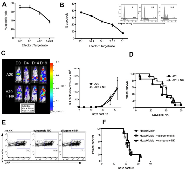
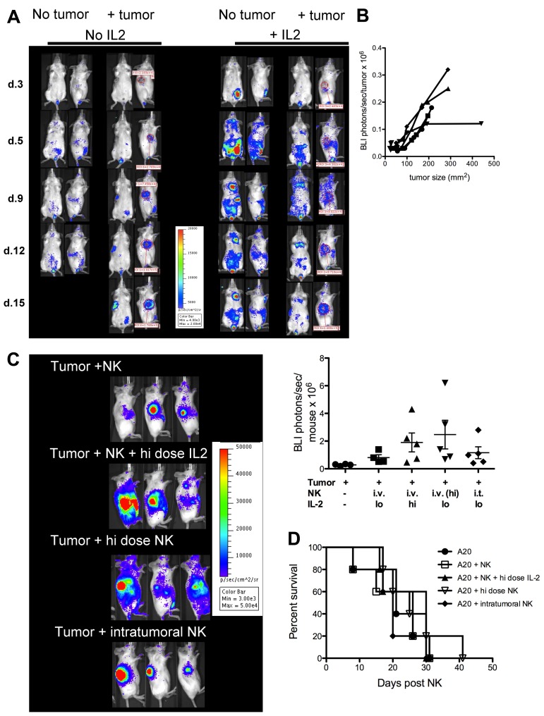
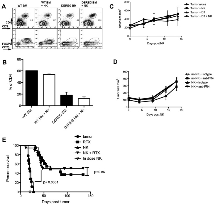
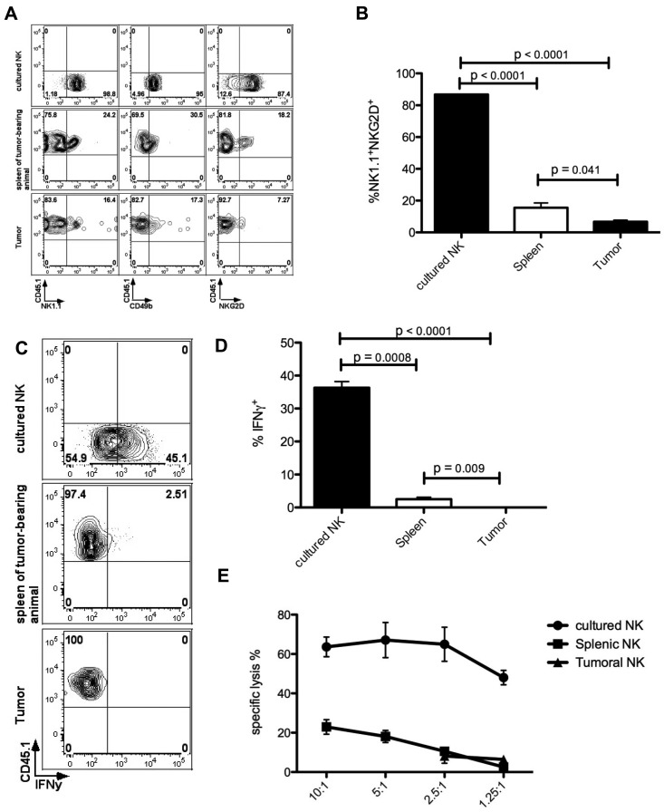
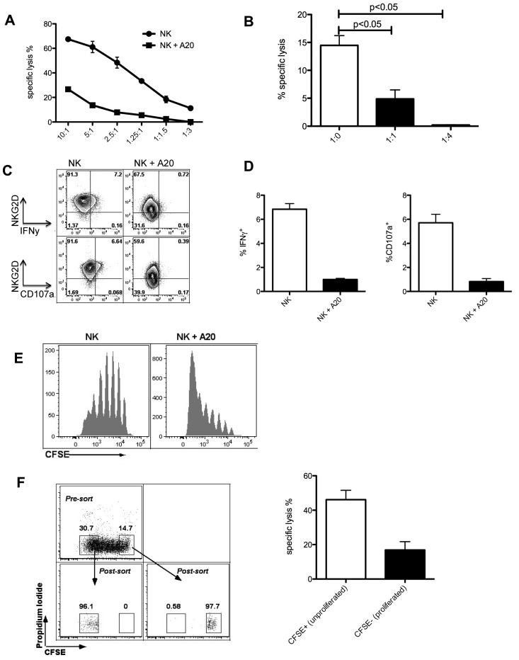
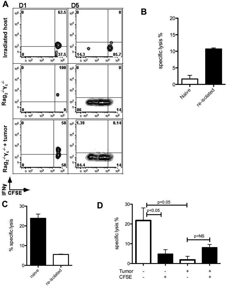
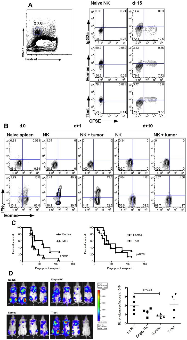
Natural killer (NK) cells are potent anti-viral and antitumor "first responders" endowed with natural cytotoxicity and cytokine production capabilities. To date, attempts to translate these promising biologic functions through the adoptive transfer of NK cells for the treatment of cancer have been of limited benefit. Here we trace the fate of adoptively transferred murine NK cells and make the surprising observation that NK cells traffic to tumor sites yet fail to control tumor growth or improve survival. This dysfunction is related to a rapid down-regulation of activating receptor expression and loss of important effector functions. Loss of interferon (IFN)γ production occurs early after transfer, whereas loss of cytotoxicity progresses with homeostatic proliferation and tumor exposure. The dysfunctional phenotype is accompanied by down-regulation of the transcription factors Eomesodermin and T-bet, and can be partially reversed by the forced overexpression of Eomesodermin. These results provide the first demonstration of NK-cell exhaustion and suggest that the NK-cell first-response capability is intrinsically limited. Further, novel approaches may be required to circumvent the described dysfunctional phenotype.


Figures

Figure 1
Figure 1
Adoptive transfer of NK cells fails to control the growth of tumor targets shown to be NK-sensitive in vitro. (A) Specific lysis of chromium labeled A20 tumor cells by IL-2–stimulated C57BL/6 NK after a 4-hour incubation. Median ± error is shown. (B) Primary AML cells (C57BL/6, H-2b) did not uptake chromium and apoptosis was detected by flow cytometric caspase activation assay after a 2-hour incubation with IL-2 stimulated Balb/c NK cells. Inset shows an example of caspase activity flow cytometry plots. Median ± error is shown. (C) Tumor burden by BLI of Balb/c mice receiving 1 × 10⁶ luciferase-expressing A20 tumor cells on day −7, followed on day 0 by TBI, BM rescue and a concurrent infusion of 0.5 × 10⁶ C57BL/6 NK cells (bottom panel) or PBS (top panel). One representative mouse from each group is shown (left), and summary statistics with n = 4 per group (right). (D) Survival of mice bearing disseminated A20 lymphoma treated with TBI and BM rescue with or without NK cells; P = .62. (E) Tumor burden by flow cytometry of BM from C57BL/6 mice 26 days after injection of 1 × 10³ gfp+ Hoxa9-Meis1 leukemia with or without 1 × 10⁶ NK cells (allogeneic FVB, H-2q; syngeneic C57BL/6, H-2b) after TBI and BM rescue. Gated on total live cells. (F) Survival of mice receiving allogeneic (FVB H-2q) or syngeneic (C57BL6 H-2b) NK cells along with Hoxa9-Meis1 leukemia; P = .93. Data are representative of at least 3 experiments with at least 4 mice per group (A,C,E), or are a composite of 2 experiments with n = 13 to 14 per group (D,F).
Figure 2
Figure 2
NK cells traffic to and accumulate within tumor sites. (A) Trafficking of C57BL/6 luc+ NK cells to A20 tumor implanted subcutaneously in Balb/c recipients. Red circles indicate tumor site and size; control groups received NK cells without tumor implantation. ROI quantification indicates photons/s/area and correlates with the number of NK cells within the site. Mice received PBS (left) or recombinant human IL-2 5 × 10⁴ units intraperitoneally every second day (right). One mouse representative of each group is shown. (B) Quantification of NK-cell accumulation by BLI in the group receiving no IL-2. NK-cells accumulate in proportion to tumor growth. Each line represents an individual mouse. Results are representative of 5 individual experiments with up to 5 mice per group. (C) NK-cell homing and accumulation on day 14 within tumor-bearing Balb/c mice receiving standard-dose (1 × 10⁶) freshly isolated C57BL/6 NK; standard-dose NK in combination with high-dose IL-2 (5 × 10⁵ units daily for 14 days); high-dose NK cells (6 × 10⁶) stimulated ex vivo in IL-2 for 5 days; or intratumoral standard-dose NK. Unless stated otherwise all groups received standard-dose IL-2 5 × 10⁴ units every second day. NK cells were derived from luciferase-transgenic animals. Three mice from each group are shown (left panel) and BLI is quantified (right panel). ANOVA P = .18 for differences in BLI between the different groups. (D). Survival of animals in (C); P = .88. Results are representative of 2 experiments containing 5 mice in each group.
Figure 3
Figure 3
NK-cell dysfunction is not rescued by depletion of regulatory T cells or recruitment of ADCC. (A-B) CD4+CD25+FoxP3+ Tregs infiltrate the tumor microenvironment. Effective depletion of Tregs can be achieved after injection of DT every second day into C57BL/6 reconstituted with DEREG or WT control BM; P = .006 for WT mice versus DEREG mice in the NK groups (2-tailed unpaired Student t test). (C) Genetic depletion of Tregs has no effect on tumor growth after NK-cell infusion; ANOVA P = .68. (D) Antibody-mediated depletion of Tregs using the anti-FR4 antibody TH6 does not impact tumor growth after NK-cell infusion. TH6 (anti-FR4) or isotype control were injected intravenously every 3 days beginning day 0 for 2 weeks; ANOVA P = .80. (E) Treatment of SCID mice bearing the human B-cell lymphoma Raji with in vitro expanded human NK cells and rituximab. Mice received tumor with isotype antibody (n = 18) or with rituximab 100 μg (n = 19), NK cells 1 × 10⁷ (n = 14), high-dose NK cells 2 × 10⁷ (n = 5), or NK 1 × 10⁷+ rituximab (n = 18). Results are representative of 2 experiments with 4 to 5 mice per group (A-D) or are a composite of 4 experiments (E).
Figure 4
Figure 4
Tumor-infiltrating NK cells down-regulate activating receptors and show impaired effector functions. (A) Purified CD3DX5+ or CD3NK1.1+ NK cells from CD45.1+ congenic donors were transferred into CD45.2+ recipients. All analyses are gated on live singlet lymphocytes. Within the spleen and tumor of tumor-bearing mice (rows 2 and 3), transferred CD45.1+ NK cells down-regulated NK1.1, NKG2D, and DX5 compared with ex vivo expanded CD45.2+ NK cells (top row). (B) Statistical comparison of events in panel A, showing marked loss of activating receptors on intratumoral NK cells compared with cultured and splenic NK cells; ANOVA with the Dunnett multiple comparison test. (C) IFNγ production is diminished within reisolated NK cells. Gating is based on nonstimulated controls (not shown). (D) Statistical comparison of events in panel C, showing loss of cytokine production on intratumoral NK cells compared with cultured or splenic NK cells; ANOVA with Dunnett multiple comparison test. (E) CD45.1+ cells were sorted from spleen or tumor and assessed for cytotoxicity against chromium-labeled A20 cells. Analyses are gated on reisolated CD45.1+ or control CD45.2+ live NK cells. Results are representative of at least 3 (A-D) or 2 (E) independent experiments with at least 3 mice per group.
Figure 5
Figure 5
NK-cell dysfunction is induced by proliferation in the presence of tumor. (A) Killing of chromium-labeled A20 cells by allogeneic C57BL/6 sorted NK that had been cultured at a 1:1 ratio with or without irradiated A20 for 5 days, in presence of IL-2 750 U/mL; effector:target ratios for the chromium assay are indicated on the x-axis. (B) Killing of chromium-labeled A20 cells by C57BL/6 NK cells that had been cultured at the indicated ratios with irradiated A20 tumor cells for 5 days, effector:target ratio for the chromium release assay was 2.5:1; P = .0009, ANOVA with Dunnett multiple comparison test. (C) Exposure to tumor leads to loss of IFNγ production and degranulation. After a 5 day exposure to irradiated A20 cells, C57BL/6 NK cells were stimulated with plate-bound anti-NK1.1 antibody and stained for IFNγ or CD107a. (D) Quantification of the events in panel C; P < .001 for IFNγ production and P < .01 for CD107a degranulation; 2-tailed unpaired Student t test. (E) Tumor coculture leads to increased NK-cell proliferation. (F) Proliferated, CFSE-low NK cells undergo more marked dysfunction than unproliferated cells. C57BL/6 NK cells were labeled with CFSE and cultured with irradiated A20 tumor cells for 4 days, then sorted on the basis of CFSE dilution; sorted cells were then cultured with chromium-labeled A20 cells at a 1:1 ratio for 18 hours; P = .03 (2-tailed unpaired Student t test). Results were done in triplicate and are representative of 3 (A,C,D) or 2 (B,E,F) experiments.
Figure 6
Figure 6
NK-cell dysfunction is induced by homeostatic proliferation. (A) IFNγ production in restimulated NK cells reisolated 1 or 5 days after in vivo transfer with or without disseminated tumor; gates were set using nonstimulated controls (not shown). Injection into Rag2−/−γc−/− recipients in addition to irradiated recipients was performed to account for the effects of radiation. (B) Killing of chromium-labeled A20 cells by freshly isolated naive NK cells or C57BL/6 CD45.1+ NK reisolated 18 hours after transfer into irradiated hosts bearing Hoxa9-Meis1 leukemia, and cultured with A20 tumor cells at a 1:1 ratio for 16 hours. Control cells were sorted CD3NK1.1+ NK cells from naive spleens; P = .0035 (2-tailed unpaired Student t test). (C) Killing of chromium-labeled A20 cells by freshly isolated naive NK cells or C57BL/6 CD45.1+ NK reisolated 17 days after transfer into irradiated hosts bearing Hoxa9-Meis1 leukemia, and cultured with A20 tumor cells indicated ratios for 16 hours. Control cells were sorted CD3NK1.1+ NK cells from naive spleens; P = .001 for difference between naive and reisolated NK cells at a 6:1 ET ratio. (D) CFSE-labeled C57BL6 NK cells were injected into Rag2−/−γc−/− recipients alone or with 5 × 10⁶ A20 tumor cells and sorted into CFSE high (unproliferated) or CFSE low (proliferated) populations; these cells were then cultured with chromium-labeled A20 cells for 12 hours at an effector:target ratio of 2:1; ANOVA with the Dunnett multiple comparison test. Results are representative of 2 to 3 experiments with 3 to 4 mice pooled per experiment.
Figure 7
Figure 7
Transcription factors Eomesodermin and T-bet are down-regulated on exposure to tumor and on proliferation. (A) Levels of Eomesodermin and T-bet fall with cell proliferation. Gates were set using isotype controls as shown in the top row for each analysis. Naive NK cells were analyzed fresh and hence were not CFSE-labeled. NK cells reisolated at D+ 15 after adoptive transfer into nontumor-bearing hosts were isolated using the expression of congenic markers, as shown in the left panel. (B) Eomes expression and IFNγ production decrease over time. NK cells were reisolated 1 or 10 days after transfer without or with 1 × 10⁵ Hoxa9-Meis1 leukemia cells. Gates are set using isotype controls, as shown in the top row. Naive spleens are depicted for comparison in the left column. (C) Sorted C57BL/6 NK cells were transduced with Eomes, T-bet, or control vector and injected with A20 cells at an E/T ratio of 5:1 into irradiated Balb/c recipients. Overexpression of Eomes, and not T-bet, leads to a significantly prolonged survival compared with control treated NK cells. (D) Tumor burden is reduced in mice receiving Eomes-transduced NK cells. Sorted C57BL/6 NK cells were transduced with Eomes, T-bet, or control vector and injected with luc+ A20 cells at a 5:1 ratio into irradiated Balb/c recipients. BLI on D+8 after injection shows a significant reduction in tumor burden among mice receiving Eomes-transduced NK cells compared with no NK cells. T-bet or control vector treated NK cells did not show a significant reduction in tumor burden. Results are representative of 3 (A) or 2 (B-D) individual experiments, or are a composite of 3 independent experiments with a total of 13 to 14 mice per group (C).

References

    1. Lundqvist A, Berg M, Smith A, Childs RW. Bortezomib treatment to Potentiate the antitumor immunity of ex-vivo expanded adoptively infused autologous natural killer cells. J Cancer. 2011;2:383–385. - PMC - PubMed
    1. Cooley S, Gada P, McKenna D, et al. Successful haploidentical hematopoietic cell engraftment using a non-myeloablative preparative regimen including natural killer (NK) cells [abstract]. Blood (ASH Annual Meeting Abstracts) 2008;112 Abstract 827.
    1. Curti A, Ruggeri L, D'Addio A, et al. Successful transfer of alloreactive haploidentical KIR ligand-mismatched natural killer cells after infusion in elderly high-risk acute myeloid leukemia patients. Blood. 2011;118(12):3273–3279. - PubMed
    1. Rubnitz JE, Inaba H, Ribeiro RC, et al. NKAML: A Pilot Study to Determine the Safety and Feasibility of Haploidentical Natural Killer Cell Transplantation in Childhood Acute Myeloid Leukemia. J Clin Oncol. 2010;28(6):955–959. - PMC - PubMed
    1. Law T, Motzer R, Mazumdar M. Phase III randomized trial of interleukin-2 with or without lymphokine-activated killer cells in the treatment of patients with advanced renal cell carcinoma. Cancer. 1995;76(5):824–832. - PubMed

Publication types

MeSH terms