. 2012 Jun 7;119(23):5467-77.
doi: 10.1182/blood-2012-01-401547. Epub 2012 Apr 27.

IKKα-mediated signaling circuitry regulates early B lymphopoiesis during hematopoiesis

Affiliations

IKKα-mediated signaling circuitry regulates early B lymphopoiesis during hematopoiesis

Mumtaz Yaseen Balkhi et al. Blood. .

Abstract

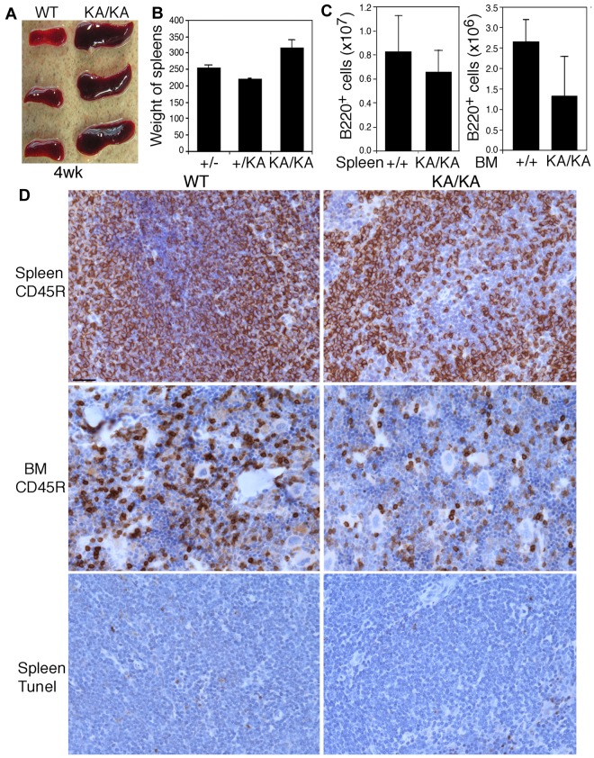
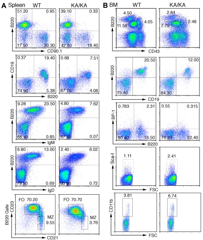
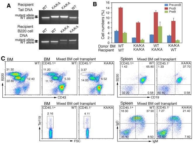
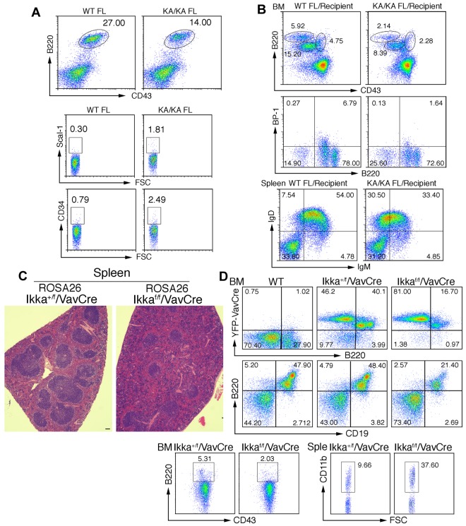
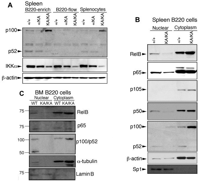
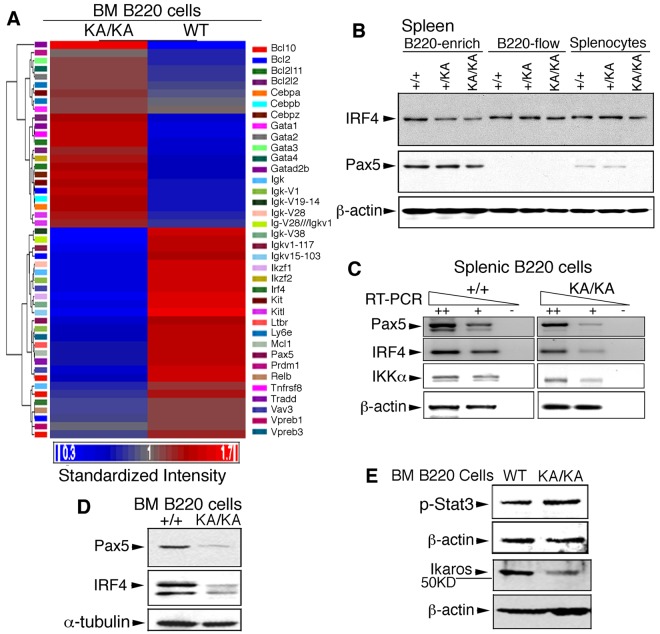
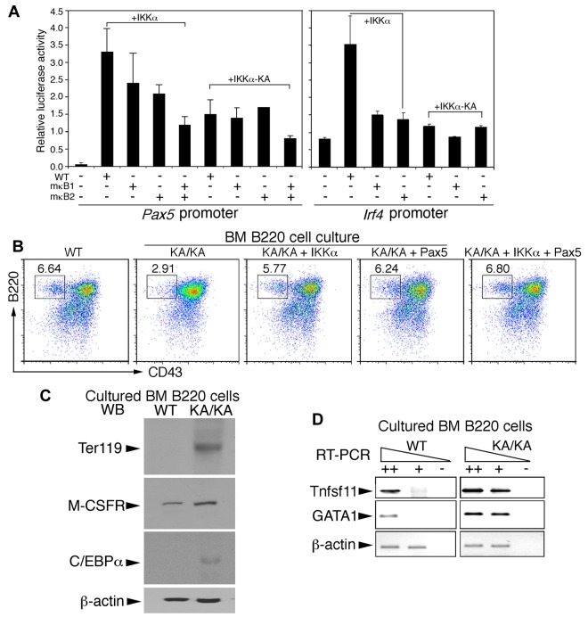
Multiple transcription factors regulate B-cell commitment, which is coordinated with myeloid-erythroid lineage differentiation. NF-κB has long been speculated to regulate early B-cell development; however, this issue remains controversial. IκB kinase-α (IKKα) is required for splenic B-cell maturation but not for BM B-cell development. In the present study, we unexpectedly found defective BM B-cell development and increased myeloid-erythroid lineages in kinase-dead IKKα (KA/KA) knock-in mice. Markedly increased cytosolic p100, an NF-κB2-inhibitory form, and reduced nuclear NF-κB p65, RelB, p50, and p52, and IKKα were observed in KA/KA splenic and BM B cells. Several B- and myeloid-erythroid-cell regulators, including Pax5, were deregulated in KA/KA BM B cells. Using fetal liver and BM congenic transplantations and deleting IKKα from early hematopoietic cells in mice, this defect was identified as being B cell-intrinsic and an early event during hematopoiesis. Reintroducing IKKα, Pax5, or combined NF-κB molecules promoted B-cell development but repressed myeloid-erythroid cell differentiation in KA/KA BM B cells. The results of the present study demonstrate that IKKα regulates B-lineage commitment via combined canonical and noncanonical NF-κB transcriptional activities to target Pax5 expression during hematopoiesis.


Figures

Figure 1
Figure 1
Reduction in the number of B cells from the spleens and BM of KA/KA mice compared with WT mice. (A-C) Spleen sizes (A), spleen weights (B), and the number of B220+ cells from the spleens and BM (C) of WT (+/+), +/KA, and KA/KA mice at 4 weeks of age. Each group contained 3 mice. The data are presented as means ± SD. (D) Comparison of B cells (CD45R) and cell death (TUNEL staining) in the spleens and BM of WT and KA/KA mice. Brown indicates immunohistochemical-stained cells with an anti-CD45R Ab or TUNEL assay; blue, nuclear countering staining. Scale bar indicates 50 μm.
Figure 2
Figure 2
Defects in splenic and BM B-cell development in KA/KA mice. (A) Analysis of the total splenic B-cell population of WT and KA/KA mice at 6 weeks of age using flow cytometry with the indicated B-cell surface markers B220+CD90.1+, B220+CD19+, B220+IgM+, B220+IgD+, CD21+CD23 (B220+ gate), and CD21loCD23hi (B220+ gate). Numbers are the percentage of the cell population; MZ, marginal zone; FO, follicular. (B) Analysis of BM cells isolated from WT and KA/KA mice at 6 weeks of age using flow cytometry with B-cell and progenitor-cell markers. A combination of markers was used to define pre-pro-B cells (B220+CD43hiCD19BP1CD24loIgM), pro-B cells (B220+CD43medBP-1+CD19+ IgM), and pre-B cells (B220hiCD43CD19+IgM).
Figure 3
Figure 3
Intrinsic B-cell defect in KA/KA BM. (A) Genotype of the tail DNA and BM B220+ cell DNA of WT and KA/KA recipient mice analyzed using PCR. (B) Statistical analysis of BM pre-pro-B, pro-B, and pre-B-cell populations of γ-irradiated recipient mice receiving BM from the donor mice as indicated. The data represent means ± SD calculated from 4 independent BM-transfer experiments. (C) Flow cytometric analysis of BM and splenic WT (CD45.1+) and KA/KA (CD45.1) B cells in the BM and spleens of irradiated mice receiving mixed WT (CD45.1+) and KA/KA (CD45.1) BM cells.
Figure 4
Figure 4
IKKα inactivation-mediated B-cell defect is an early event. (A) Flow cytometric analysis of B-cell and progenitor profiles in the FL of embryonic day 13.5 WT and KA/KA mouse embryos. (B) Analyses of BM and splenic B-cell profiles of irradiated Rag−/− mice (Recipients) receiving WT and KA/KA FL (embryonic day 13.5) cells using flow cytometry with B220+CD43+, B220+BP-1+, and IgD+IgM+ markers. (C) Histopathology of H&E-stained spleens from Ikkα+/f/VavCre/ROSA26 and Ikkαf/f/VavCre/ROSA26 mice. Scale bar indicates 50 μm. (D) Analyses of the BM and splenic (Sple) B-cell profiles of Ikkα+/f/VavCre/ROSA26 and Ikkαf/f/VavCre/ROSA26 mice using flow cytometry with B220+, B220+CD43+, B220+CD19+, and CD11b+ markers.
Figure 5
Figure 5
Reduced activity of canonical and noncanonical NF-κB components in the splenic and BM B cells of KA/KA mice. (A) Western blot showing the indicated protein levels in splenic cells. B220-enrich indicates column-purified B220+ cells; B220-flow, non-B cells that passed through the column; splenocytes, total cell lysate from whole spleen; β-actin, protein-loading control. (B-C) p65 (RelA), RelB, p100/52, and p105/p50 expression levels in nuclear and cytoplasmic fractions of spleen and BM B220+ cells of WT (+/+) and KA/KA mice. Lamin B and sp1 were used as controls for the purity of nuclear extracts.
Figure 6
Figure 6
IKKα inactivation deregulates the expression of genes involved in BM B-cell development. (A) Heat map and hierarchical cluster analysis expression of 36 genes (supplemental Table 2) in WT and KA/KA BM B220+ cells, as identified using microarray analysis. Red and blue indicate up-regulated and down-regulated genes, respectively. Standardized intensity is indicated by the fold values (Log2) of gene expression in WT and KA/KA BM B cells. (B) Western blot showing IRF4 and Pax5 levels in B220+-enriched cells, B220-flow, and splenocyte lysates (see Figure 5A). β-actin, protein-loading control (same as in Figure 5A). (C) Expression of Pax5, IRF4, and IKKα analyzed with RT-PCR on serially diluted total RNA isolated from the BM B220+ cells of WT and KA/KA mice. ++, +, cDNA dilution; -, negative control; β-actin, PCR control. (D) IRF4 and Pax5 levels in the BM B220+ cells of WT (+/+) and KA/KA mice detected with Western blot. α-tubulin, protein-loading control. (E) Stat3 activity (p-Stat3) and Ikaros level in the BM B220+ cells of WT and KA/KA mice. β-actin, protein-loading control.
Figure 7
Figure 7
IKKα and combined noncanonical and canonical NF-κB regulate Pax5 and Irf4 promoter activities and BM B-cell differentiation. (A) The activities of Pax5, Irf4, mutant Pax5, and mutant Irf4 promoters containing mutations within κB1 (mκB1) or κB2 (mκB2) in pGL3b luciferase reporter vectors were examined in Raji B cells using the luciferase reporter assay. +IKKα indicates coexpression of IKKα and promoter plasmids; +IKKα-KA, coexpression of IKKα-KA and promoter plasmids. (B) Flow cytometric analyses of B220+ BM B cells in RPMI 1640 medium supplemented with 10% FBS for 3 days after reintroduction of IKKα, IKKα-KA, Pax5, and IKKα+Pax5 plasmids into BM B cells using AMAXA nucleofection. (C) Levels of Ter119, M-CSFR, and C/EBPα in cultured WT and KA/KA BM B cells were detected with Western blotting. β-actin, protein-loading control. (D) RT-PCR showing expression levels of Tnfsf11 and GAGA1 in cultured WT and KA/KA BM B cells. ++, +, cDNA dilution; -, negative control; β-actin, PCR control.

References

    1. Busslinger M. Transcriptional control of early B cell development. Annu Rev Immunol. 2004;22:55–79. - PubMed
    1. Kaisho T, Takeda K, Tsujimura T, et al. IkappaB kinase alpha is essential for mature B cell development and function. J Exp Med. 2001;193(4):417–426. - PMC - PubMed
    1. Senftleben U, Cao Y, Xiao G, et al. Activation by IKKalpha of a second, evolutionary conserved, NF-kappa B signaling pathway. Science. 2001;293(5534):1495–1499. - PubMed
    1. Horwitz BH, Scott ML, Cherry SR, Bronson RT, Baltimore D. Failure of lymphopoiesis after adoptive transfer of NF-kappaB-deficient fetal liver cells. Immunity. 1997;6(6):765–772. - PubMed
    1. Guo F, Tanzer S, Busslinger M, Weih F. Lack of nuclear factor-kappa B2/p100 causes a RelB-dependent block in early B lymphopoiesis. Blood. 2008;112(3):551–559. - PubMed

Publication types

MeSH terms