A nomogram for individualized estimation of survival among patients with brain metastasis
- PMID: 22544733
- PMCID: PMC3379797
- DOI: 10.1093/neuonc/nos087
A nomogram for individualized estimation of survival among patients with brain metastasis
Abstract
Purpose: An estimated 24%-45% of patients with cancer develop brain metastases. Individualized estimation of survival for patients with brain metastasis could be useful for counseling patients on clinical outcomes and prognosis.
Methods: De-identified data for 2367 patients with brain metastasis from 7 Radiation Therapy Oncology Group randomized trials were used to develop and internally validate a prognostic nomogram for estimation of survival among patients with brain metastasis. The prognostic accuracy for survival from 3 statistical approaches (Cox proportional hazards regression, recursive partitioning analysis [RPA], and random survival forests) was calculated using the concordance index. A nomogram for 12-month, 6-month, and median survival was generated using the most parsimonious model.
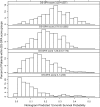
Results: The majority of patients had lung cancer, controlled primary disease, no surgery, Karnofsky performance score (KPS) ≥ 70, and multiple brain metastases and were in RPA class II or had a Diagnosis-Specific Graded Prognostic Assessment (DS-GPA) score of 1.25-2.5. The overall median survival was 136 days (95% confidence interval, 126-144 days). We built the nomogram using the model that included primary site and histology, status of primary disease, metastatic spread, age, KPS, and number of brain lesions. The potential use of individualized survival estimation is demonstrated by showing the heterogeneous distribution of the individual 12-month survival in each RPA class or DS-GPA score group.
Conclusion: Our nomogram provides individualized estimates of survival, compared with current RPA and DS-GPA group estimates. This tool could be useful for counseling patients with respect to clinical outcomes and prognosis.
Figures


References
-
- Eichler AF, Loeffler JS. Multidisciplinary management of brain metastases. Oncologist. 2007;12:884–898. doi:10.1634/theoncologist.12-7-884. - DOI - PubMed
-
- Sawaya R, Bindal RK, Lang FF, Abi-Said D. Metastatic brain tumors. In: Kaye AH, Laws ER, editors. Brain Tumors An Encyclopedic Approach. 2nd ed. London: Churchill Livingstone; 2001. pp. 999–1026.
-
- Barnholtz-Sloan JS, Sloan AE, Davis FG, Vigneau FD, Lai P, Sawaya RE. Incidence proportions of brain metastases in patients diagnosed (1973 to 2001) in the Metropolitan Detroit Cancer Surveillance System. J Clin Oncol. 2004;22:2865–2872. doi:10.1200/JCO.2004.12.149. - DOI - PubMed
-
- Gaspar LE, Scott C, Murray K, Curran W. Validation of the RTOG recursive partitioning analysis (RPA) classification for brain metastases. Int J Radiat Oncol Biol Phys. 2000;47:1001–1006. doi:10.1016/S0360-3016(00)00547-2. - DOI - PubMed
-
- Gaspar L, Scott C, Rotman M, et al. Recursive partitioning analysis (RPA) of prognostic factors in three Radiation Therapy Oncology Group (RTOG) brain metastases trials. Int J Radiat Oncol Biol Phys. 1997;37:745–751. doi:10.1016/S0360-3016(96)00619-0. - DOI - PubMed
Publication types
MeSH terms
Grants and funding
LinkOut - more resources
Full Text Sources
Other Literature Sources
Medical

