. 2012 Jun 15;287(25):21406-15.
doi: 10.1074/jbc.M112.370213. Epub 2012 Apr 27.

TGD1, -2, and -3 proteins involved in lipid trafficking form ATP-binding cassette (ABC) transporter with multiple substrate-binding proteins

Affiliations

TGD1, -2, and -3 proteins involved in lipid trafficking form ATP-binding cassette (ABC) transporter with multiple substrate-binding proteins

Rebecca L Roston et al. J Biol Chem. .

Abstract

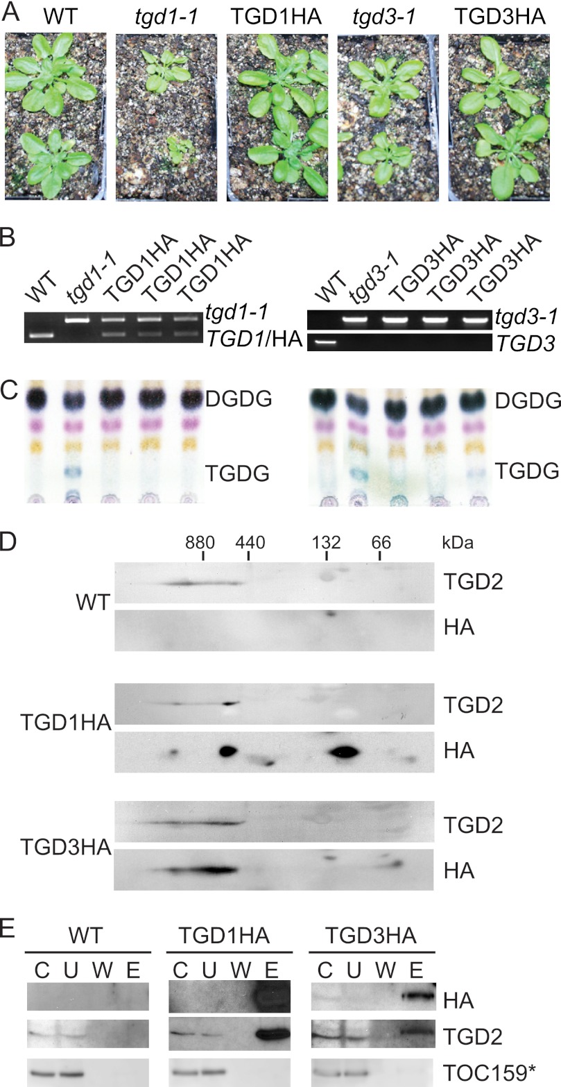
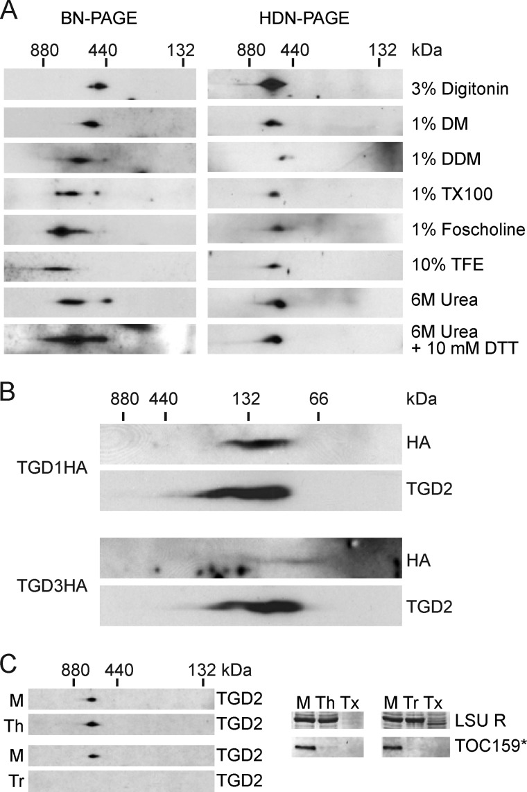
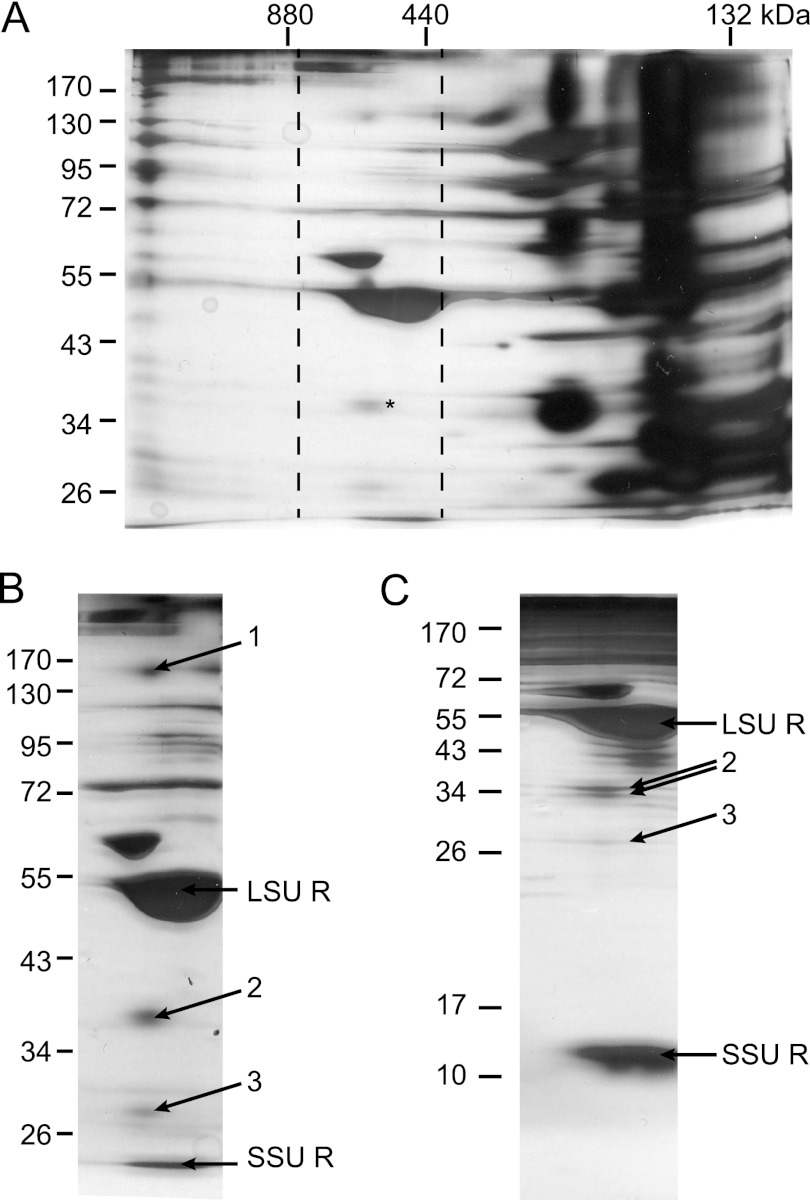
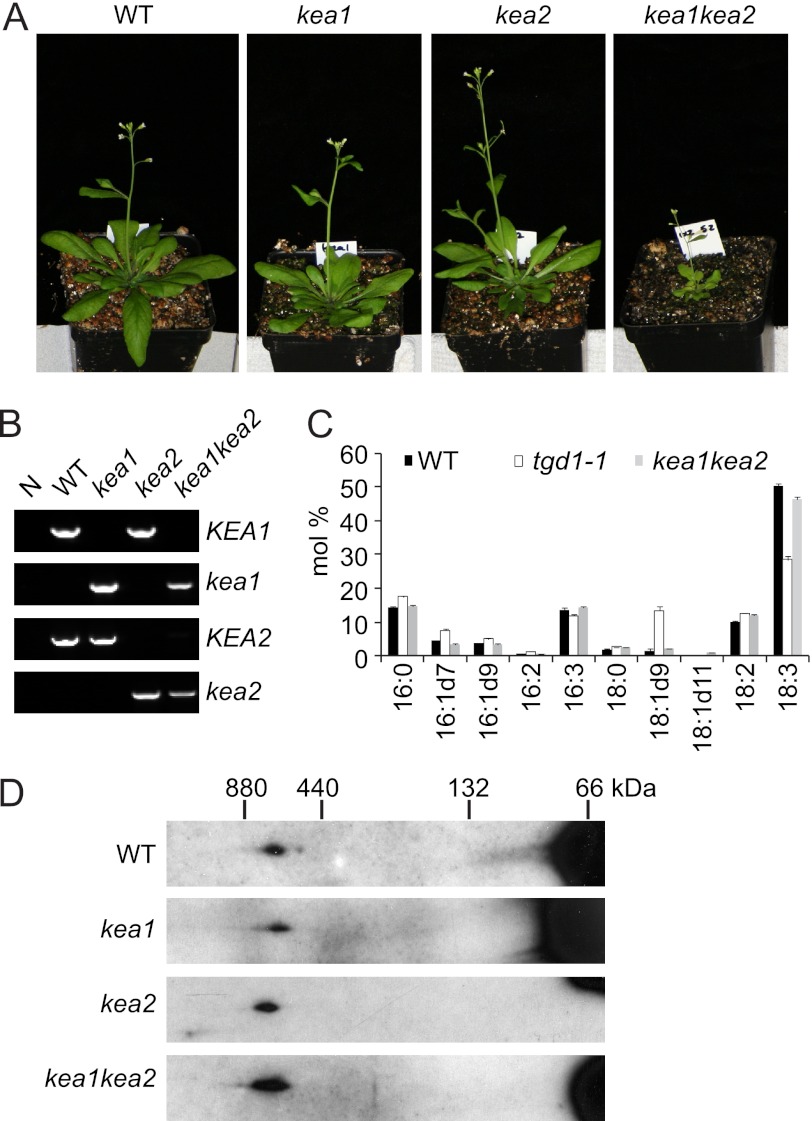
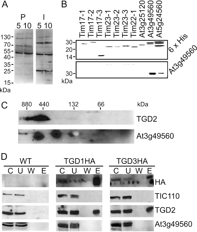
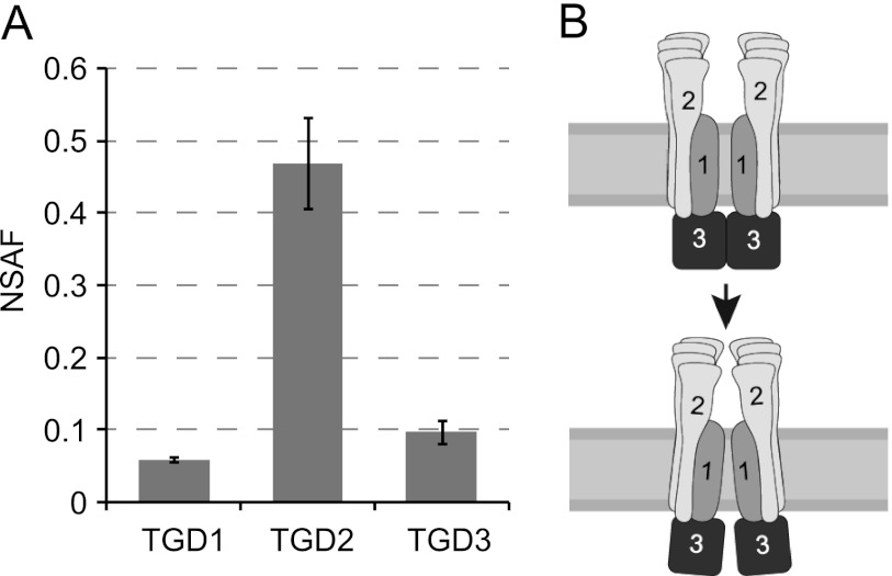
Members of the ATP-binding cassette (ABC) transporter family are essential proteins in species as diverse as archaea and humans. Their domain architecture has remained relatively fixed across these species, with rare exceptions. Here, we show one exception to be the trigalactosyldiacylglycerol 1, 2, and 3 (TGD1, -2, and -3) putative lipid transporter located at the chloroplast inner envelope membrane. TGD2 was previously shown to be in a complex of >500 kDa. We demonstrate that this complex also contains TGD1 and -3 and is very stable because it cannot be broken down by gentle denaturants to form a "core" complex similar in size to standard ABC transporters. The complex was purified from Pisum sativum (pea) chloroplast envelopes by native gel electrophoresis and examined by mass spectrometry. Identified proteins besides TGD1, -2, or -3 included a potassium efflux antiporter and a TIM17/22/23 family protein, but these were shown to be in separate high molecular mass complexes. Quantification of the complex components explained the size of the complex because 8-12 copies of the substrate-binding protein (TGD2) were found per functional transporter.


Figures

FIGURE 1.
FIGURE 1.
TGD1, -2, and -3 associate in a large complex. A, 28-day-old Arabidopsis are shown, with genotypes as labeled above (WT indicates wild-type Arabidopsis). TGD1HA or TGD3HA labels indicate plants homozygous for the tgd1-1 or tgd3-1 alleles, which also express TGD1HA or TGD3HA under the control of the 35S promoter, respectively. B, genotyping of plants as labeled showing the presence of the mutant tgd1-1 or tgd3-1 alleles and lack of the endogenous TGD1 or TGD3 alleles. C, α-naphthol-stained, thin layer chromatogram of lipids isolated from Arabidopsis of genotypes given at the top. Digalactosyldiacylglycerol (DGDG) and trigalactosyldiacylglycerol (TGDG) are indicated. D, immunoblots detecting proteins indicated at the right of 20 μg of 1% dodecylmaltoside-solubilized, chlorophyll-equivalent chloroplasts isolated from Arabidopsis of genotypes indicated at the left. Protein complexes were separated in the first dimension on a 4–14% BN-PAGE (marker at top) and then denatured and run on a 12% SDS-PAGE in the second dimension. E, immunoblots detecting proteins listed at the right (TOC159* indicates the 86-kDa fragment of TOC159) of an immunoprecipitation using HA antiserum of solubilized Arabidopsis chloroplasts, genotypes given at the top. C, chloroplast starting material; U, unbound fraction; W, final wash; E, eluate.
FIGURE 2.
FIGURE 2.
The stable TGD1, -2, and -3 complex is present in the chloroplast inner envelope. A, anti-TGD2 immunoblots of two-dimensional gels in which the first dimension is a 4–10% BN-PAGE (left) or HDN-PAGE (right) and the second dimension is a 12% SDS-PAGE. Isolated wild-type Arabidopsis chloroplasts (equivalent to 20 μg of chlorophyll) were solubilized with reagents listed at the right (DM, decylmaltoside; TX100, Triton X-100; TFE, trifluoroethanol). In the case of trifluoroethanol and urea samples, 1% DDM was also present. B, immunoblots detecting proteins indicated at the right, similar to the HDN-PAGEs in A, except the gradient was 4–14%, and chloroplasts isolated from Arabidopsis of genotypes indicated at the left were solubilized in 1% SDS. C, anti-TGD2 immunoblots similar to the HDN-PAGE solubilized with DDM in A, except prior to solubilization the chloroplasts were mock-treated (M), thermolysin-treated (Th), or trypsin-treated (Tr). Immunoblots of single-dimension SDS-PAGEs show digestion of the 86-kDa fragment of TOC159 (TOC159*), and Coomassie Brilliant Blue stain shows protection of the large subunit of Rubisco (LSU R). Tx, trypsin treatment with Triton X-100 present.
FIGURE 3.
FIGURE 3.
The TGD complex of pea co-migrates with another high molecular mass protein. A, isolated pea chloroplast envelopes solubilized in 3% digitonin and separated by 4–14% HDN-PAGE in the first dimension and 10% SDS-PAGE in the second and then visualized by silver staining. Dashed lines mark the selection of gels displayed in B and C. An asterisk marks location of TGD2. B, as in A, with arrows showing bands submitted for mass spectrometry analysis. The large and small subunits of Rubisco are also labeled (LSU R and SSU R, respectively). C, as in B, except 15% SDS-PAGE in the second dimension. These data were not used in final analysis because band 1 could not be separated under these conditions.
FIGURE 4.
FIGURE 4.
Potassium efflux antiporters are not part of the TGD complex. A, 35-day-old Arabidopsis are shown with genotypes labeled above. B, genomic DNA extracted from genotypes listed above (N, no-DNA control) was genotyped using PCR with primers recognizing alleles indicated at the right (KEA1 is the wild-type KEA1 allele, and kea1 is the T-DNA insertion into KEA1; KEA2 and kea2 for the KEA2 allele are designated accordingly). C, total fatty acids of lipids converted to fatty acid methylesters and quantified by gas chromatography for genotypes listed above. The S.D. value of three biological replicates is shown (error bars). D, dodecylmaltoside-solubilized leaf proteins from Arabidopsis genotypes listed at the left were separated in the first dimension by 4–14% HDN-PAGE and in the second dimension by 12% SDS-PAGE and then immunoblotted and detected with TGD2 antiserum.
FIGURE 5.
FIGURE 5.
TIM family proteins are not part of the TGD complex. A, immunoblots of isolated chloroplast protein equivalent to 5 or 10 μg of chlorophyll were probed with At3g49560 antiserum (I) or its preimmune serum (P), as indicated at the top. B, antiserum raised against proteins encoded by At3g49560 was tested for specificity against the preprotein and amino acid transporter family. Full-length recombinant proteins for Tim17-1 (At1g20350), Tim17-2 (At2g37410), Tim17-3 (At5g11690), Tim23-1 (At1g17530), Tim23-2 (At1g72750), Tim23-3 (At3g04800), Tim22-1 (At1g18320/At3g10110), At3g25120, At3g49560, and At5g24650 (indicated at the top) were detected with antiserum recognizing proteins indicated at the right. C, chloroplasts isolated from wild-type Arabidopsis were separated by 4–14% HDN-PAGE in the first dimension and 12% SDS-PAGE in the second and then immunoblotted and detected with antisera recognizing proteins indicated at the right. D, immunoprecipitations of HA-tagged proteins, similar to those in Fig. 1E, were immunoblotted and probed with antisera recognizing proteins indicated at the right. C, chloroplast starting material; U, unbound fraction; W, final wash; E, eluate.
FIGURE 6.
FIGURE 6.
There are 8–10 copies of TGD2 per functional transporter. A, calculated normalized spectral abundance factors (NSAF) are displayed with S.D. (error bars) for TGD1, -2, and -3. B, model of the TGD1, -2, and -3 complex suggests that changes in TGD1 geometry due to ATPase activity by TGD3 could be shared by multiple TGD2 polypeptides.

References

    1. Albers S. V., Koning S. M., Konings W. N., Driessen A. J. (2004) Insights into ABC transport in archaea. J. Bioenerg. Biomembr. 36, 5–15 - PubMed
    1. Higgins C. F. (1992) ABC transporters. From microorganisms to man. Annu. Rev. Cell Biol. 8, 67–113 - PubMed
    1. Saier M. H., Jr. (2000) A functional-phylogenetic classification system for transmembrane solute transporters. Microbiol. Mol. Biol. Rev. 64, 354–411 - PMC - PubMed
    1. Hollenstein K., Dawson R. J., Locher K. P. (2007) Structure and mechanism of ABC transporter proteins. Curr. Opin. Struct. Biol. 17, 412–418 - PubMed
    1. van der Heide T., Poolman B. (2002) ABC transporters. One, two, or four extracytoplasmic substrate-binding sites? EMBO Rep. 3, 938–943 - PMC - PubMed

Publication types

MeSH terms