. 2012 Jun 15;287(25):21461-70.
doi: 10.1074/jbc.M112.344358. Epub 2012 Apr 27.

Crystal structure of DnaK protein complexed with nucleotide exchange factor GrpE in DnaK chaperone system: insight into intermolecular communication

Affiliations

Crystal structure of DnaK protein complexed with nucleotide exchange factor GrpE in DnaK chaperone system: insight into intermolecular communication

Ching-Chung Wu et al. J Biol Chem. .

Abstract

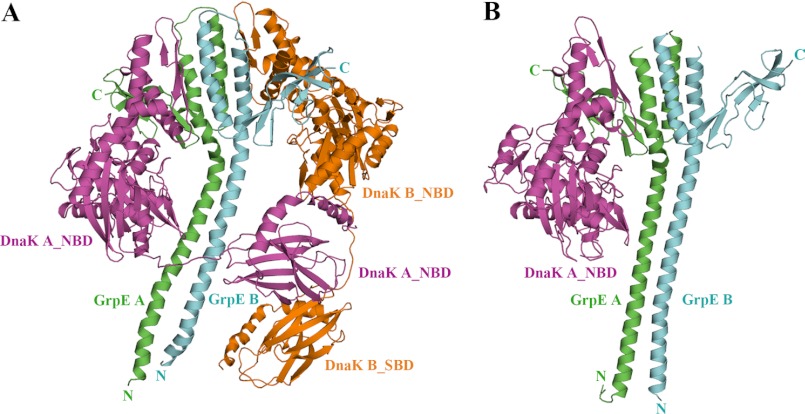
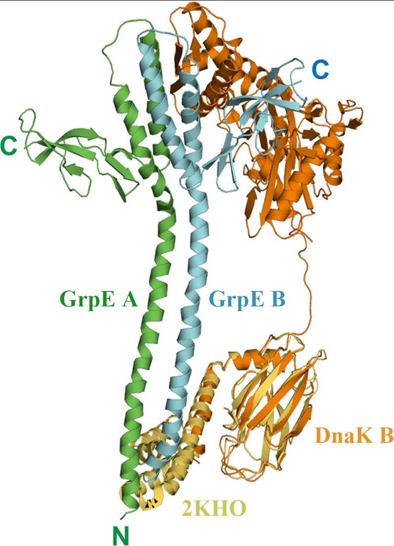
The conserved, ATP-dependent bacterial DnaK chaperones process client substrates with the aid of the co-chaperones DnaJ and GrpE. However, in the absence of structural information, how these proteins communicate with each other cannot be fully delineated. For the study reported here, we solved the crystal structure of a full-length Geobacillus kaustophilus HTA426 GrpE homodimer in complex with a nearly full-length G. kaustophilus HTA426 DnaK that contains the interdomain linker (acting as a pseudo-substrate), and the N-terminal nucleotide-binding and C-terminal substrate-binding domains at 4.1-Å resolution. Each complex contains two DnaKs and two GrpEs, which is a stoichiometry that has not been found before. The long N-terminal GrpE α-helices stabilize the linker of DnaK in the complex. Furthermore, interactions between the DnaK substrate-binding domain and the N-terminal disordered region of GrpE may accelerate substrate release from DnaK. These findings provide molecular mechanisms for substrate binding, processing, and release during the Hsp70 chaperone cycle.


Figures

FIGURE 1.
FIGURE 1.
Overall structure of the GkDnaK-GkGrpE complex and comparison with the EcoDnaK_NBD-EcoGrpE complex. A, ribbon diagram of the GkDnaK-GkGrpE complex showing the GkGrpE homodimer (green, GrpE A; cyan, GrpE B) bound by two GkDnaK molecules (magenta, DnaK A; orange, DnaK B). B, ribbon diagram of the EcoDnaK_NBD-EcoGrpE complex (PDB code 1DKG). The color coding is the same as in A.
FIGURE 2.
FIGURE 2.
Stoichiometry of GkDnaK with GkGrpE homodimer. A, gel filtration analysis of GkDnaK-GkGrpE complex. The elution profiles colored in blue and in red represent the complex protein solution before crystallization and the complex solution dissolved from protein crystals, respectively. The x-axis and y-axis represent the elution volume and optical intensity by UV spectrometer at 280 nm, respectively. Inset, SDS-PAGE analysis of GkDnaK-GkGrpE complex dissolved from crystals. B, interprotein interactions GkDnaK and GkGrpE performed by increasing concentrations of GkGrpE protein at different molar ratios (lanes 3-8) as indicated with control proteins (protein sample do not contain partner protein; lanes 1 and 2). The individual bands corresponding to GkDnaK and GkGrpE were observed to disappear at equimolar ratio (lane 5). A further increase in GkGrpE concentration (lanes 6-8) over GkDnaK does not result in further binding indicating that the interaction follows 2:2 stoichiometry, not 2:1 as speculated earlier in Gk species. C, upper panels, heat release/s after addition of aliquots of the GkGrpE homodimer into a calorimetry cell containing GkDnaK or GkDnaK_NBD. Lower panels, integrated binding isotherms (black circles, derived from upper panel) and experimental fits (solid red lines) to a single-site model. The best-fit molar-binding stoichiometry values are 1.59 and 1.88 for GkGrpE with GkDnaK and GkDnaK_NBD, respectively.
FIGURE 3.
FIGURE 3.
Structural comparison of DnaK molecules in different nucleotide-binding states from different species. A, NBDs of GkDnaK in the ADP/Mg2+/Pi state (magenta; PDB code 2V7Y) and the nucleotide-free state (present study; green, DnaK A; cyan, DnaK B) superimposed to show the large scale movement of the SBDs. B, Superpositioning of GkDnaK_NBD in the ADP/Mg2+/Pi state (magenta) and EcoDnaK_NBD (cyan) and GkDnaK_NBD (green), both in nucleotide-free state. The bound nucleotide (ADP) in GkDnaK_NBD is shown as a stick model. The dashed lines indicate the angle(s) (°) of the open nucleotide-binding pockets among the different structures. C, interaction of the DnaK_NBD subdomain IIB with the GrpE four-helix bundle or the C-terminal β-sheet domain. GkGrpE A and B are shown as green and cyan cylinders, respectively. Subdomains IIB of nucleotide-free GkDnaK_NBD and EcoDnaK_NBD are colored in magenta and orange. Magenta and orange asterisks indicate major interacting regions between GrpE and the GkDnaK and EcoDnaK subdomains IIB, respectively.
FIGURE 4.
FIGURE 4.
Structural comparison of GrpEs from different species. A, front and orthogonal views of GkGrpE (green) aligned with TthGrpE (orange; PDB code 3A6M) and EcoGrpE (blue; PDB code 1DKG). Large shifts in the orientations and positions occur mainly in the long N-terminal α-helices. B, superpositioning of the long GrpE N-terminal α-helices showing substantially more curvature for the four-helix bundle in GkGrpE (46.7°, green, chain A) compared with four-helix bundle in EcoGrpE (24.1°, blue) and TthGrpE (9.4°, yellow). Red dashed lines indicate central axes.
FIGURE 5.
FIGURE 5.
Topology of GkGrpE homodimer. Stereo view of GkGrpE initial experimental electron density map around the linker region connecting α-helices at the four-helix bundle domain. Chains A and B of GkGrpE are represented in green and blue, respectively. The observed electron density is shown in blue and purple mesh at 1.0σ and 2.0σ, respectively, and the residues involved in connecting the helices at four-helix bundle domain are represented with legends.
FIGURE 6.
FIGURE 6.
Interactions between GkDnaK and the GkGrpE homodimer. A, possible side chain interactions between GkDnaK A and the GkGrpE homodimer. GkGrpE A (green) and B (blue) are shown as cylinders with side chains. GkDnaK residues in the NBD (magenta), interdomain linker (yellow), and SBD (orange) are shown in stick models. Possible hydrogen bonds are shown as dashed lines. B, complementation assay for DnaK. Serial dilutions of fresh E. coli cultures were spotted onto agar containing LB and incubated at 37 °C or 42 °C overnight. Empty vector and full-length DnaK (EcoDnaK_FL and GkDnaK_FL) vectors were used as negative and positive controls, respectively. Dilution factors for the E. coli cultures are labeled over the panels.
FIGURE 7.
FIGURE 7.
Molecular modeling between GkDnaK and GkGrpE homodimer. Ribbon diagram describes the modeling result of GkDnaK (orange, DnaK B) aligned with the complete EcoDnaK_SBD structure (yellow; PDB code 2KHO). The mimic GkDnaK full-length model shows the possible interaction between the extended α-helical lid domain of GkDnaK and the N-terminal disordered and long helical region of GkGrpE dimer (green, GrpE A; cyan, GrpE B).

References

    1. Mayer M. P., Bukau B. (2005) Hsp70 chaperones: cellular functions and molecular mechanism. Cell. Mol. life Sci. 62, 670–684 - PMC - PubMed
    1. Meimaridou E., Gooljar S. B., Chapple J. P. (2009) From hatching to dispatching: the multiple cellular roles of the Hsp70 molecular chaperone machinery. J. Mol. Endocrinol. 42, 1–9 - PubMed
    1. Hartl F. U. (1996) Molecular chaperones in cellular protein folding. Nature 381, 571–579 - PubMed
    1. Hartl F. U., Hayer-Hartl M. (2002) Molecular chaperones in the cytosol: from nascent chain to folded protein. Science 295, 1852–1858 - PubMed
    1. Rohde M., Daugaard M., Jensen M. H., Helin K., Nylandsted J., Jäättelä M. (2005) Members of the heat-shock protein 70 family promote cancer cell growth by distinct mechanisms. Genes Dev. 19, 570–582 - PMC - PubMed

Publication types

MeSH terms

Associated data

LinkOut - more resources