. 2012 Nov;56(5):1902-12.
doi: 10.1002/hep.25817. Epub 2012 Oct 9.

CD11b(+) Gr1(+) bone marrow cells ameliorate liver fibrosis by producing interleukin-10 in mice

Affiliations

CD11b(+) Gr1(+) bone marrow cells ameliorate liver fibrosis by producing interleukin-10 in mice

Yang-Gun Suh et al. Hepatology. 2012 Nov.

Abstract

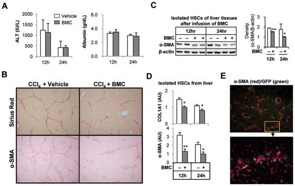
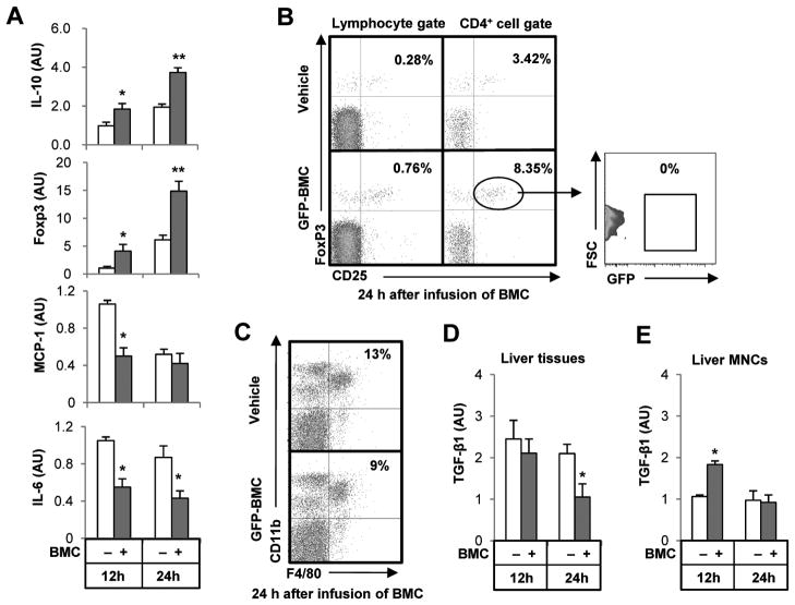
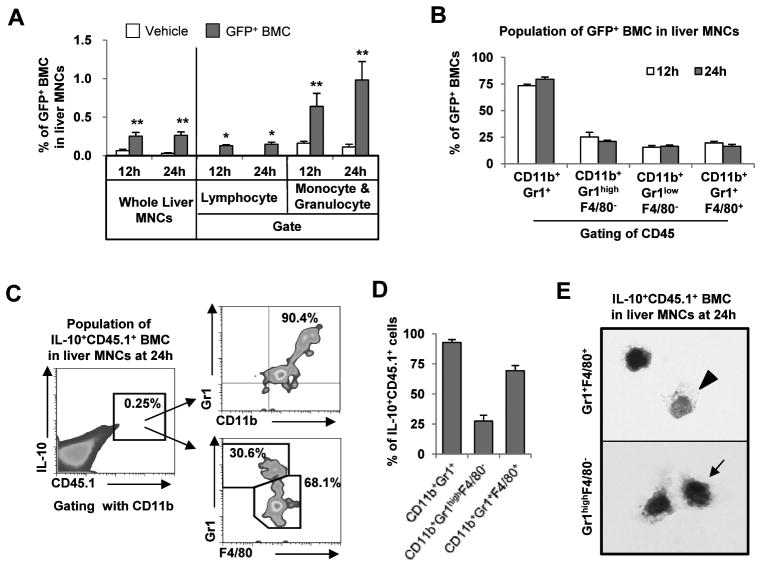
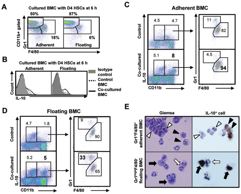
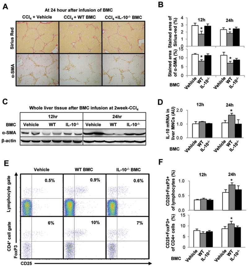
Clinical trials and animal models suggest that infusion of bone marrow cells (BMCs) is effective therapy for liver fibrosis, but the underlying mechanisms are obscure, especially those associated with early effects of BMCs. Here, we analyzed the early impact of BMC infusion and identified the subsets of BMCs showing antifibrotic effects in mice with carbon tetrachloride-induced liver fibrosis. An interaction between BMCs and activated hepatic stellate cells (HSCs) was investigated using an in vitro coculturing system. Within 24 hours, infused BMCs were in close contact with activated HSCs, which was associated with reduced liver fibrosis, enhanced hepatic expression of interleukin (IL)-10, and expanded regulatory T cells but decreased macrophage infiltration in the liver at 24 hours after BMC infusion. In contrast, IL-10-deficient (IL-10(-/-) ) BMCs failed to reproduce these effects in fibrotic livers. Intriguingly, in isolated cells, CD11b(+) Gr1(high) F4/80(-) and CD11b(+) Gr1(+) F4/80(+) BMCs expressed more IL-10 after coculturing with activated HSCs, leading to suppressed expression of collagen and α-smooth muscle actin in HSCs. Moreover, these effects were either enhanced or abrogated, respectively, when BMCs were cocultured with IL-6(-/-) and retinaldehyde dehydrogenase 1(-/-) HSCs. Similar to murine data, human BMCs expressed more IL-10 after coculturing with human HSC lines (LX-2 or hTERT), and serum IL-10 levels were significantly elevated in patients with liver cirrhosis after autologous BMC infusion.

Conclusion: Activated HSCs increase IL-10 expression in BMCs (CD11b(+) Gr1(high) F4/80(-) and CD11b(+) Gr1(+) F4/80(+) cells), which in turn ameliorates liver fibrosis. Our findings could enhance the design of BMC therapy for liver fibrosis.


Figures

Fig. 1
Fig. 1
Infused BMC ameliorates CCl4-induced liver fibrosis in mice. Mice with 2-week CCl4–induced liver fibrosis were sacrificed at 12 or 24 hours after infusion of BMC of GFP+ mice via tail vein. (A) Serum chemistry was conducted for ALT and albumin. (B) Representative Sirius red or α-SMA staining of liver tissue at 24 hours after BMC infusion (original magnification, ×100). (C) Isolated HSCs of liver tissues were subjected to Western blotting. (D) α-SMA and COL1A1 mRNA levels in isolated HSCs were determined by quantitative RT-PCR (qRT-PCR). (E) After staining with α-SMA antibody, frozen liver sections were observed under the fluorescent microscopy (original magnification, upper panels ×100; lower panels ×400). Data are expressed as the mean ± SEM (at least 8 ~ 12 mice per group). *P < 0.05, **P < 0.01 in comparison with the corresponding controls.
Fig. 2
Fig. 2
Infused BMC increase IL-10 expression and CD4+CD25+Foxp3+ Treg while decreasing the expression of IL-6 and MCP-1, and reducing F4/80+CD11b+ cells in fibrotic liver. Mice with 2-weeks of CCl4–induced liver fibrosis were sacrificed at 12 or 24 hours after infusion of BMC of GFP+ mice via tail vein, and then liver MNCs and tissues were isolated or collected. (A) Isolated liver MNCs were subjected to qRT-PCR. (B, C) CD4+CD25+Foxp3+ Tregs and F4/80+CD11b+ macrophages in liver MNCs were analyzed at 24 hour after infusion of BMC. (D, E) TGF-β1 mRNA levels of whole liver tissues and isolated liver MNCs were determined by qRT-PCR. Data are expressed as the mean ± SEM (at least 8 mice per group). *P < 0.05, **P < 0.01 in comparison with the corresponding controls.
Fig. 3
Fig. 3
Infused BMC subtypes CD11b+Gr1+F4/80+ and CD11b+Gr1highF4/80 play important roles in IL-10 production in fibrotic livers of recipient mice. Mice with 2-week CCl4–induced liver fibrosis were sacrificed at 12 or 24 hours after infusion of BMC of GFP+ or CD45.1+ mice via tail vein. Liver MNCs were isolated and subjected to FACS analyses. (A) The percentages of infused BMC (GFP+) to total liver MNCs were analyzed. (B) The cellular markers of GFP+ BMC were analyzed using antibodies to CD45, CD11b, Gr1 and F4/80. (C, D) Intracellular cytokine staining of IL-10 was performed and analyzed for IL-10+CD45.1+ BMC of isolate liver MNCs. (E) Among IL-10+CD45.1+ BMC of isolate liver MNCs, CD11b+Gr1+F4/80+ and CD11b+Gr1highF4/80 BMCs were sorted and stained with Giemsa respectively (Original magnification, ×2000). Data are expressed as the mean ± SEM (at least 5 ~ 10 mice per group). *P < 0.05, **P < 0.01 in comparison with the corresponding controls.
Fig. 4
Fig. 4
Co-culturing with HSCs enhances IL-10 expression by BMC, which suppresses activation of D4 HSCs. (A) Isolated HSCs from healthy mice were cultured on plastic dish for 4 days (D4 HSCs). Then freshly isolated BMC were co-cultured with activated D4 HSCs for 6 to 24 hours (HSCs:BMC = 1:10). (B) IL-10 mRNA levels in adherent and floating BMC of mice were determined by qRT-PCR. (C) After co-culturing, α-SMA and COL1A1 mRNA levels of D4 HSCs of mice were determined by qRT-PCR. (D) Human HSCs (LX-2 and hTERT cell lines) were co-cultured with human BMC of HBV patients for 6 to 24 hours after 6 hour-starvation of human HSCs. IL-10 mRNA levels in floating human BMC were determined by qRT-PCR. (E) IL-10 levels of sera in HBV patients (n=15) with cirrhosis were measured at various time points and analyzed after autologous BMC infusion. (F) After autologous BMC infusion, IL-10 levels in blood were analyzed in the effective patients (n=10) of all patients (n=15) showing the improvement of Child-Pugh scoring and albumin levels. Data are expressed as the mean ± SEM via 3 ~ 5 independent experiments. *P < 0.05, **P < 0.01 in comparison with the corresponding controls.
Fig. 5
Fig. 5
CD11b+Gr1highF4/80 and CD11b+Gr1+F4/80+ BMC enhance IL-10 expression after co-culturing with D4 HSCs. BMC isolated from normal mice were co-cultured with D4 HSCs. (A) Adherent and floating BMC were analyzed using antibodies to Gr1 and F4/80 after co-culturing with D4 HSCs for 6 hours. (B) IL-10 positive BMC increased in both adherent and floating BMC after co-culturing. (C, D) IL-10 positive BMC were analyzed using antibodies to IL-10, CD11b, Gr1 and F4/80. (E) Co-cultured BMC (CD11b+Gr1highF4/80 and CD11b+Gr1+F4/80+) were sorted using FACS Aria and then stained with Giemsa or intracellular cytokine staining of IL-10 were performed after further culturing with monensin for additional 18 hours (original magnification, ×1200). IL-10 positive cells were indicated as black arrowhead or black arrow, and IL-10 negative cells were shown as white arrowhead or white arrow. Data are expressed as the mean ± SEM via 5 independent experiments. *P < 0.05, **P < 0.01 in comparison with the corresponding controls.
Fig. 6
Fig. 6
Depletion of IL-10 gene in BMC abrogates the anti-fibrotic effects of BMC via down regulation of IL-10 expression and Treg induction in liver MNCs of recipient mice. Mice with 2-week CCl4-induced liver fibrosis were infused with vehicle, isolated whole BMC of control WT or IL-10−/− mice through tail vein. Mice were sacrificed at 12 and 24 hours after BMC infusion. (A, B) To estimate liver fibrosis and HSC activation, sections of liver tissues were stained with Sirius red and α-SMA antibody. The stained areas of Sirius red and α-SMA antibody were calculated with image analyzer program. (C) Whole liver tissues were subjected to Western blotting. (D) IL-10 mRNA levels in liver MNCs were determined qRT-PCR analyses. (E, F) Intrahepatic CD4+CD25+FoxP3+ Treg increased in the mice treated with WT BMC but not in IL-10−/− BMC at 24 hour. Data are expressed as the mean ± SEM (at least 8 ~ 10 mice per group). *P < 0.05 in comparison with the corresponding controls.
Fig. 7
Fig. 7
IL-10 expression of BMC is promoted or suppressed by retinoic acid and IL-6 of HSCs, respectively. Isolated HSCs from WT, IL-6−/−, IL-10−/− and RALDH1−/− mice were cultured on plastic plates for 4 days (D4 HSCs). Then freshly isolated WT BMC from control mice were co-cultured with activated D4 HSCs for 6 to 24 hours (HSCs:BMC = 1:10) or cultured alone as control. (A, B) After co-culturing, IL-10 expressions in the adherent and floating BMC were analyzed using qRT-PCR. RALDH1−/− mice with 2-weekCCl4-induced liver fibrosis were infused with isolated whole BMC of WT mice through tail vein and then were sacrificed at 24 h after infusion of BMC. (C) Representative Sirius red staining of liver tissue (original magnification, ×100). (D) There were no significant changes in the population of Gr1+CD11b+ and F4/80+CD11b+ cells in liver, and intrahepatic CD4+CD25+FoxP3+ Treg did not increase in RALDH1−/− mice after infusion of WT BMC at 24 hours. (E) An integrated depiction of anti-fibrotic roles of infused BMC after interaction with HSCs. First, the interaction between infused BMC and HSCs is crucial for IL-10 production in infused BMC (especially CD11b+Gr1+F4/80+ and CD11b+Gr1highF4/80 MDSC-like cells). Second, retinoic acid and IL-6 from activated HSCs stimulate and inhibit respectively the production of IL-10 by infused BMC. Third, IL-10 from infused BMC of donor mice is essential for the induction of Treg in recipient mice, which then suppresses activated HSCs via direct (IL-10 production) and indirect (Treg induction) effects of infused BMC. Data are expressed as the mean ± SEM via 3 independent experiments or using 8 mice per group. *P < 0.05, **P < 0.01 in comparison with the corresponding controls.

References

    1. Petersen BE, Bowen WC, Patrene KD, Mars WM, Sullivan AK, Murase N, et al. Bone marrow as a potential source of hepatic oval cells. Science. 1999;284:1168–1170. - PubMed
    1. Sakaida I, Terai S, Yamamoto N, Aoyama K, Ishikawa T, Nishina H, et al. Transplantation of bone marrow cells reduces CCl4-induced liver fibrosis in mice. Hepatology. 2004;40:1304–1311. - PubMed
    1. Terai S, Ishikawa T, Omori K, Aoyama K, Marumoto Y, Urata Y, et al. Improved liver function in patients with liver cirrhosis after autologous bone marrow cell infusion therapy. Stem Cells. 2006;24:2292–2298. - PubMed
    1. Biermann H, Pietz B, Dreier R, Schmid KW, Sorg C, Sunderkotter C. Murine leukocytes with ring-shaped nuclei include granulocytes, monocytes, and their precursors. J Leukoc Biol. 1999;65:217–231. - PubMed
    1. Hernandez-Gea V, Friedman SL. Pathogenesis of liver fibrosis. Annu Rev Pathol. 2011;6:425–456. - PubMed

Publication types

MeSH terms