

In vivo clearance of alpha-1 acid glycoprotein is influenced by the extent of its N-linked glycosylation and by its interaction with the vessel wall
- PMID: 22545002
- PMCID: PMC3321579
- DOI: 10.1155/2012/292730
In vivo clearance of alpha-1 acid glycoprotein is influenced by the extent of its N-linked glycosylation and by its interaction with the vessel wall
Abstract
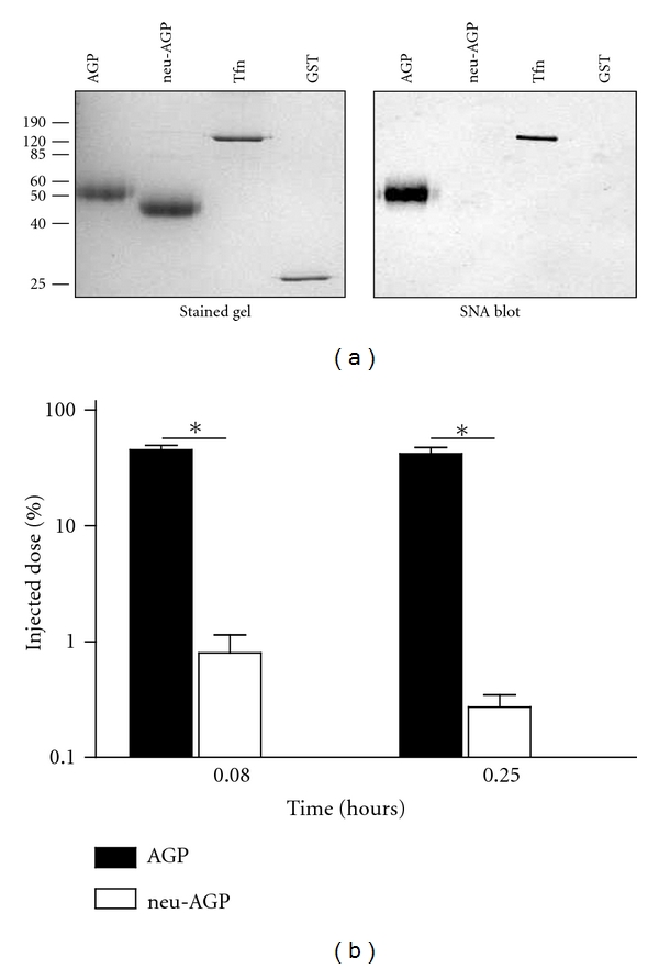
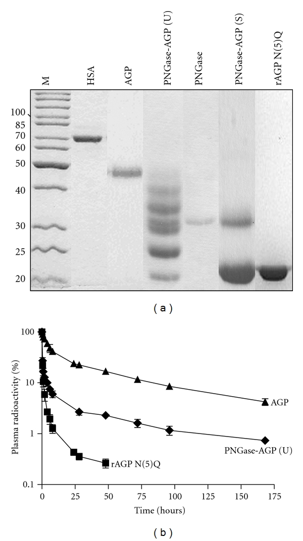
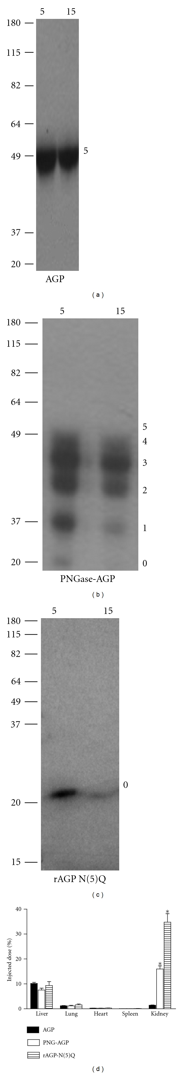
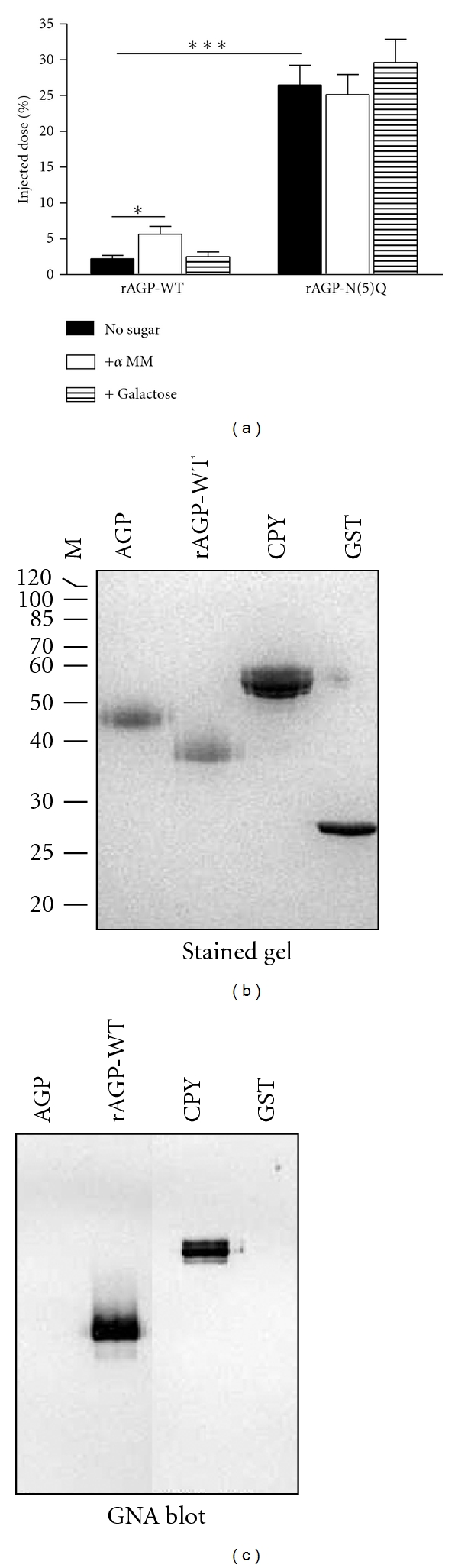
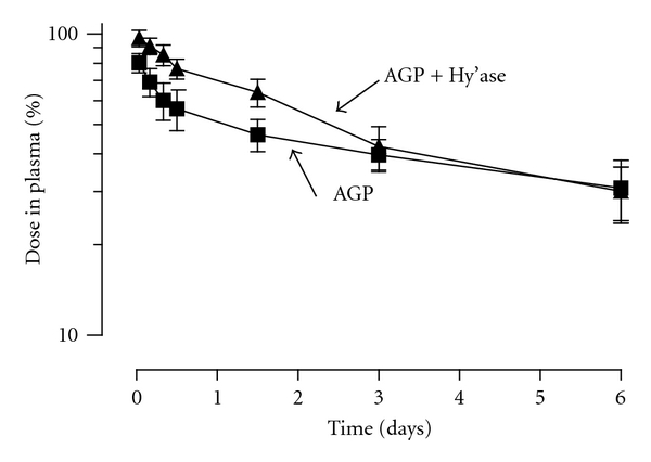
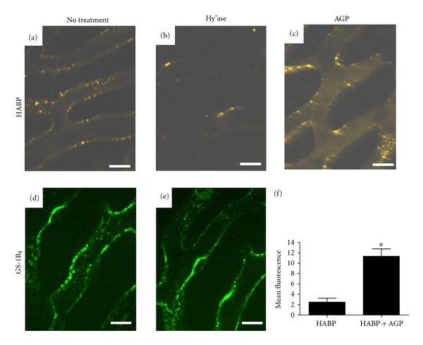
Alpha-1 acid glycoprotein (AGP) is a highly glycosylated plasma protein that exerts vasoprotective effects. We hypothesized that AGP's N-linked glycans govern its rate of clearance from the circulation, and followed the disappearance of different forms of radiolabeled human AGP from the plasma of rabbits and mice. Enzymatic deglycosylation of human plasma-derived AGP (pdAGP) by Peptide: N-Glycosidase F yielded a mixture of differentially deglycosylated forms (PNGase-AGP), while the introduction of five Asn to Gln mutations in recombinant Pichia pastoris-derived AGP (rAGP-N(5)Q) eliminated N-linked glycosylation. PNGase-AGP was cleared from the rabbit circulation 9-fold, and rAGP-N(5)Q, 46-fold more rapidly than pdAGP, primarily via a renal route. Pichia pastoris-derived wild-type rAGP differed from pdAGP in expressing mannose-terminated glycans, and, like neuraminidase-treated pdAGP, was more rapidly removed from the rabbit circulation than rAGP-N(5)Q. Systemic hyaluronidase treatment of mice transiently decreased pdAGP clearance. AGP administration to mice reduced vascular binding of hyaluronic acid binding protein in the liver microcirculation and increased its plasma levels. Our results support a critical role of N-linked glycosylation of AGP in regulating its in vivo clearance and an influence of a hyaluronidase-sensitive component of the vessel wall on its transendothelial passage.
Figures
Similar articles
-
Pichia pastoris-produced mucin-type fusion proteins with multivalent O-glycan substitution as targeting molecules for mannose-specific receptors of the immune system.Glycobiology. 2011 Aug;21(8):1071-86. doi: 10.1093/glycob/cwr046. Epub 2011 Apr 7. Glycobiology. 2011. PMID: 21474492
-
Highly efficient production of peptides: N-glycosidase F for N-glycomics analysis.Protein Expr Purif. 2014 May;97:17-22. doi: 10.1016/j.pep.2014.02.004. Epub 2014 Feb 26. Protein Expr Purif. 2014. PMID: 24582822
-
Characterization of the hepatic cellular uptake of α(1) -acid glycoprotein (AGP), part 1: a peptide moiety of human AGP is recognized by the hemoglobin β-chain on mouse liver parenchymal cells.J Pharm Sci. 2012 Apr;101(4):1599-606. doi: 10.1002/jps.22804. Epub 2011 Nov 11. J Pharm Sci. 2012. PMID: 22081433
-
The acute phase protein alpha1-acid glycoprotein: a model for altered glycosylation during diseases.Curr Protein Pept Sci. 2007 Feb;8(1):91-108. doi: 10.2174/138920307779941497. Curr Protein Pept Sci. 2007. PMID: 17305563 Review.
-
Carbohydrate composition and immunomodulatory activity of different glycoforms of alpha1-acid glycoprotein.Glycoconj J. 1997 Aug;14(5):631-8. doi: 10.1023/a:1018544711767. Glycoconj J. 1997. PMID: 9298696 Review.
Cited by
-
Distinguishing Benign and Malignant Thyroid Nodules and Identifying Lymph Node Metastasis in Papillary Thyroid Cancer by Plasma N-Glycomics.Front Endocrinol (Lausanne). 2021 Jun 25;12:692910. doi: 10.3389/fendo.2021.692910. eCollection 2021. Front Endocrinol (Lausanne). 2021. PMID: 34248851 Free PMC article.
-
Physiologically Based Pharmacokinetic Model of Brain Delivery of Plasma Protein Bound Drugs.Pharm Res. 2023 Mar;40(3):661-674. doi: 10.1007/s11095-023-03484-2. Epub 2023 Feb 24. Pharm Res. 2023. PMID: 36829100 Free PMC article.
-
Serum Linkage-Specific Sialylation Changes Are Potential Biomarkers for Monitoring and Predicting the Recurrence of Papillary Thyroid Cancer Following Thyroidectomy.Front Endocrinol (Lausanne). 2022 Apr 29;13:858325. doi: 10.3389/fendo.2022.858325. eCollection 2022. Front Endocrinol (Lausanne). 2022. PMID: 35574008 Free PMC article.
-
Validation of diagnostic and predictive biomarkers for hereditary angioedema via plasma N-glycomics.Clin Transl Allergy. 2021 Dec;11(10):e12090. doi: 10.1002/clt2.12090. Clin Transl Allergy. 2021. PMID: 34962719 Free PMC article.
-
Pharmacokinetic and Pharmacodynamic Considerations for Drugs Binding to Alpha-1-Acid Glycoprotein.Pharm Res. 2018 Dec 28;36(2):30. doi: 10.1007/s11095-018-2551-x. Pharm Res. 2018. PMID: 30593605 Free PMC article. Review.
References
-
- Fournier T, Medjoubi-N N, Porquet D. Alpha-1-acid glycoprotein. Biochimica et Biophysica Acta. 2000;1482(1-2):157–171. - PubMed
-
- Kremer JMH, Wilting J, Janssen LHM. Drug binding to human alpha-1-acid glycoprotein in health and disease. Pharmacological Reviews. 1988;40(1):1–47. - PubMed
-
- Ricca GA, Hamilton RW, McLean JW. Rat α1-acid glycoprotein mRNA. Cloning of double-stranded cDNA and kinetics of induction of mRNA levels following acute inflammation. Journal of Biological Chemistry. 1981;256(20):10362–10368. - PubMed
-
- Cooper R, Papaconstantinou J. Evidence for the existence of multiple α1-acid glycoprotein genes in the mouse. Journal of Biological Chemistry. 1986;261(4):1849–1853. - PubMed
MeSH terms
Substances
LinkOut - more resources
Full Text Sources
