. 2012 Feb;7(1):79-91.
doi: 10.2217/clp.11.70.

The juvenile Batten disease protein, CLN3, and its role in regulating anterograde and retrograde post-Golgi trafficking

Affiliations

The juvenile Batten disease protein, CLN3, and its role in regulating anterograde and retrograde post-Golgi trafficking

Susan L Cotman et al. Clin Lipidol. 2012 Feb.

Abstract

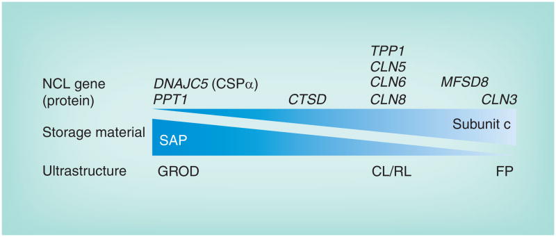
Loss-of-function mutations in CLN3 are responsible for juvenile-onset neuronal ceroid lipofuscinosis (JNCL), or Batten disease, which is an incurable lysosomal disease that manifests with vision loss, followed by seizures and progressive neurodegeneration, robbing children of motor skills, speech and cognition, and eventually leading to death in the second or third decade of life. Emerging clinical evidence points to JNCL pathology outside of the CNS, including the cardiovascular system. The CLN3 gene encodes an unusual transmembrane protein, CLN3 or battenin, whose elusive function has been the subject of intense study for more than 10 years. Owing to the detailed characterization of a large number of disease models, our knowledge of CLN3 protein function is finally coming into focus. This review will describe the most current understanding of CLN3 structure, function and dysfunction in JNCL.


Figures

Figure 1
Figure 1. The neuronal ceroid lipofuscinosis disease spectrum
Lysosomal storage material across the different genetic forms of NCL is heterogeneous and features one or more characteristic ultrastructural profiles. GRODs contain mostly SAPs A and D, and this is the most prominent profile of the storage material when PPT1 is mutated, although sometimes this is mixed with other profiles [16]. On the other end of the spectrum, CLN3 is mostly associated with FP profile storage material, although other profiles may also be present [16]. A number of genes (TPP1, CLN5, CLN6 and CLN8) that are typically linked with NCL onset in the late-infantile years are most associated with CL and RL profiles. CTSD mutations in humans typically cause a rare, severe congenic form of NCL for which there is limited patient information regarding storage material profiles [3]. However, Ctsd deficiency in the mouse leads to both GROD and FP profiles [90]. Finally, GROD storage material was the predominant finding in patients with DNAJC5 mutations. Whether or not there is a relationship between the different ultrastructural profiles of the storage material is unclear. CL: Curvilinear; FP: Fingerprint; GROD: Granular osmiophilic deposit; NCL: Neuronal ceroid lipofuscinosis; RL: Rectilinear; SAP: Saposin.
Figure 2
Figure 2. CLN3 topology and juvenile-onset neuronal ceroid lipofuscinosis mutations
The predicted topology of CLN3 is depicted. Sites for post-translational modifications are shown (blue forked lines represent N-glycosylation, zigzag represents prenylation and dotted line represents a possible cleavage site following prenylation), and point mutations are marked by red (missense) and yellow (nonsense) colored circles. A reported polymorphism at residue 404 (H/R) is shown (dark blue). Residues E295 and Q352 are both red and yellow as they are sites for both missense and nonsense mutations. A residue that has been associated with slower disease, in compound heterozygosity with the common 1.02-kb deletion mutation. A residue that has been associated with slower disease in homozygosity.
Figure 3
Figure 3. The CLN3 protein mediates anterograde and retrograde trafficking in a pathway connecting the Golgi network, endosomes, autophagosomes, lysosomes and plasma membrane
Upon synthesis in the endoplasmic reticulum and glycosylation in the Golgi, CLN3 has several potential fates that may in part be mediated by compartment-specific interacting proteins. The sorting of CLN3 into post-Golgi transport vesicles may be directed by adaptor proteins AP-1 and AP-3 in clathrin-coated membrane buds from the trans-Golgi network. A significant proportion of CLN3 then enters late endosomes, which undergo acidification to form lysosomes. A portion of the lysosomal pool fuses with autophagosomes, double membrane structures that may be derived from the Golgi (as shown) or other sources, including the endoplasmic reticulum and endosomes (not shown). CLN3 may interact with a complex of autophagy proteins including Atg3 and Atg7, which catalyze the addition of phosphatidylethanolamine to LC3 to form LC3-II, a proteolipid that assembles into the inner and outer membranes of the nascent phagophore and promotes its extension to a mature autophagosome. An undetermined fraction of CLN3 may be directed from transport vesicles to the plasma membrane or from late endosomes to the plasma membrane, where it may interact with components of the membrane cytoskeleton such as nonmuscle myosin-IIB and fodrin, which links plasma membrane proteins such as the Na+/K+-ATPase to the actin cytoskeletion. Whether CLN3 interacts with these proteins as an endosomal protein (as shown) or as a plasma membrane protein is unclear. The possible interaction of CLN3 with AP-2, the adaptor protein involved in clathrin-mediated endocytosis of plasma membrane proteins (labeled as cargo receptors), is less clear. A portion of CLN3 may facilitate the transport of sphingolipids, including GalCer, from the trans Golgi to the plasma membrane. Recent evidence in yeast models suggests that CLN3 or its yeast ortholog Btn1 (shown) is involved in retrograde transport from the endocytic compartment to the trans-Golgi network. This may be mediated indirectly by assembly of the SNARE protein Sed5, which must be phosphorylated to direct retrograde transport. Sed5 is either directly or indirectly phosphorylated by the palmitoylated endosome- and vacuole-associated casein kinase Yck3. It was speculated that Btn1p may exert a desaturase activity on the palmitoyl moiety of Yck3 and thereby promote its activation of Sed5. CLN3 interactors are shown in red text. GalCer: Galactosylceramide.

References

    1. Kohlschutter A, Schulz A. Towards understanding the neuronal ceroid lipofuscinoses. Brain Dev. 2009;31(7):499–502. - PubMed
    1. Jalanko A, Braulke T. Neuronal ceroid lipofuscinoses. Biochim Biophys Acta. 2009;1793(4):697–709. - PubMed
    1. Mole S, Williams R, Goebel H. The Neuronal Ceroid Lipofuscinoses (Batten Disease) 2. Oxford University Press; UK: 2011. Comprehensive review of the clinicopathologic features of neuronal ceroid lipofuscinosis (NCL), the spectrum of mutations in each of the NCL genes known at the time of its publication, the genetic models that have been developed for each form of the disease and the proposed functions of each of the proteins encoded by the NCL genes.
    1. Mole S, Williams R, Goebel H. Correlations between genotype, ultrastructural morphology and clinical phenotype in the neuronal ceroid lipofuscinoses. Neurogenetics. 2005;6(3):107–126. - PubMed
    1. Chattopadhyay S, Ito M, Cooper J, et al. An autoantibody inhibitory to glutamic acid decarboxylase in the neurodegenerative disorder Batten disease. Hum Mol Genet. 2002;11(12):1421–1431. - PubMed

Websites

    1. SCOP: Structural Classification of Proteins, 1.75 release. 2009 Jun; http://scop.mrc-lmb.cam.ac.uk/scop.
    1. The Wellcome Trust. Sanger Institute. Pfam 26.0. 2011 Nov; http://pfam.sanger.ac.uk.
    1. NCL Resource – A gateway for Batten disease. www.ucl.ac.uk/ncl.
    1. UCSC Genome Bioinformatics. http://genome.ucsc.edu.
    1. Gene expression omnibus. www.ncbi.nlm.nih.gov/geo/query/acc.cgi?acc=GSE24368.

LinkOut - more resources