

Construction of vascular tissues with macro-porous nano-fibrous scaffolds and smooth muscle cells enriched from differentiated embryonic stem cells
- PMID: 22545119
- PMCID: PMC3335865
- DOI: 10.1371/journal.pone.0035580
Construction of vascular tissues with macro-porous nano-fibrous scaffolds and smooth muscle cells enriched from differentiated embryonic stem cells
Abstract
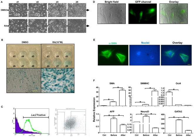
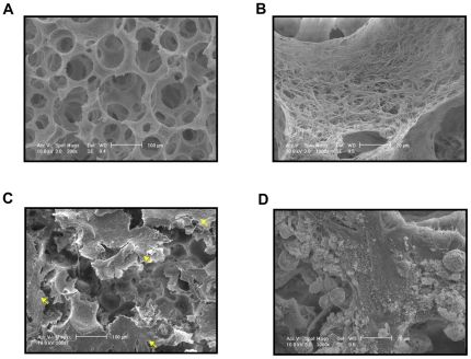
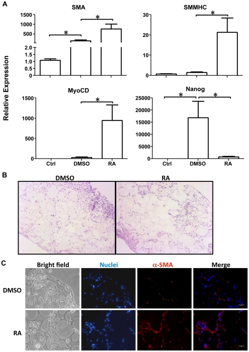
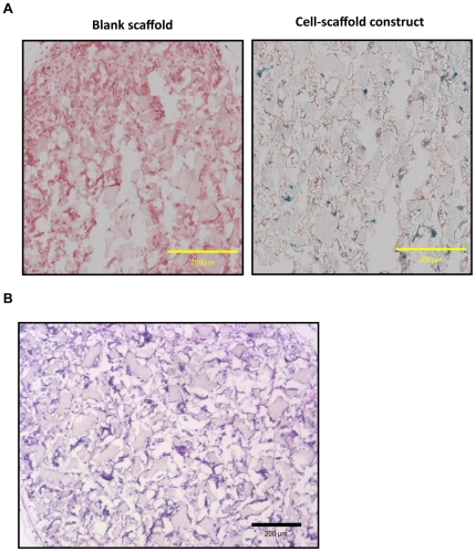
Vascular smooth muscle cells (SMCs) have been broadly used for constructing tissue-engineered blood vessels. However, the availability of mature SMCs from donors or patients is very limited. Derivation of SMCs by differentiating embryonic stem cells (ESCs) has been reported, but not widely utilized in vascular tissue engineering due to low induction efficiency and, hence, low SMC purity. To address these problems, SMCs were enriched from retinoic acid induced mouse ESCs with LacZ genetic labeling under the control of SM22α promoter as the positive sorting marker in the present study. The sorted SMCs were characterized and then cultured on three-dimensional macro-porous nano-fibrous scaffolds in vitro or implanted subcutaneously into nude mice after being seeded on the scaffolds. Our data showed that the LacZ staining, which reflected the corresponding SMC marker SM22α expression level, was efficient as a positive selection marker to dramatically enrich SMCs and eliminate other cell types. After the sorted cells were seeded into the three-dimensional nano-fibrous scaffolds, continuous retinoic acid treatment further enhanced the SMC marker gene expression level while inhibited pluripotent maker gene expression level during the in vitro culture. Meanwhile, after being implanted subcutaneously into nude mice, the implanted cells maintained the positive LacZ staining within the constructs and no teratoma formation was observed. In conclusion, our results demonstrated the potential of SMCs derived from ESCs as a promising cell source for therapeutic vascular tissue engineering and disease model applications.
Conflict of interest statement
Figures
Similar articles
-
Engineering vascular tissue with functional smooth muscle cells derived from human iPS cells and nanofibrous scaffolds.Biomaterials. 2014 Oct;35(32):8960-9. doi: 10.1016/j.biomaterials.2014.07.011. Epub 2014 Jul 29. Biomaterials. 2014. PMID: 25085858 Free PMC article.
-
Derivation of smooth muscle cells with neural crest origin from human induced pluripotent stem cells.Cells Tissues Organs. 2012;195(1-2):5-14. doi: 10.1159/000331412. Epub 2011 Oct 14. Cells Tissues Organs. 2012. PMID: 22005509 Free PMC article.
-
Three-dimensional growth of iPS cell-derived smooth muscle cells on nanofibrous scaffolds.Biomaterials. 2011 Jul;32(19):4369-75. doi: 10.1016/j.biomaterials.2011.02.049. Epub 2011 Mar 24. Biomaterials. 2011. PMID: 21439638 Free PMC article.
-
Stem cell-derived vasculature: A potent and multidimensional technology for basic research, disease modeling, and tissue engineering.Biochem Biophys Res Commun. 2016 May 6;473(3):733-42. doi: 10.1016/j.bbrc.2015.09.127. Epub 2015 Sep 30. Biochem Biophys Res Commun. 2016. PMID: 26427871 Free PMC article. Review.
-
Embryonic origins of human vascular smooth muscle cells: implications for in vitro modeling and clinical application.Cell Mol Life Sci. 2014 Jun;71(12):2271-88. doi: 10.1007/s00018-013-1554-3. Epub 2014 Jan 18. Cell Mol Life Sci. 2014. PMID: 24442477 Free PMC article. Review.
Cited by
-
Fabrication of tissue-engineered vascular grafts with stem cells and stem cell-derived vascular cells.Expert Opin Biol Ther. 2016;16(3):317-30. doi: 10.1517/14712598.2016.1118460. Epub 2015 Dec 8. Expert Opin Biol Ther. 2016. PMID: 26560995 Free PMC article. Review.
-
Biomaterials and stem cells for tissue engineering.Expert Opin Biol Ther. 2013 Apr;13(4):527-40. doi: 10.1517/14712598.2013.756468. Epub 2013 Jan 17. Expert Opin Biol Ther. 2013. PMID: 23327471 Free PMC article. Review.
-
Biomimetic tubular scaffold with heparin conjugation for rapid degradation in in situ regeneration of a small diameter neoartery.Biomaterials. 2021 Jul;274:120874. doi: 10.1016/j.biomaterials.2021.120874. Epub 2021 May 12. Biomaterials. 2021. PMID: 34051629 Free PMC article.
-
Porous nanofibrous poly(L-lactic acid) scaffolds supporting cardiovascular progenitor cells for cardiac tissue engineering.Acta Biomater. 2015 Oct;26:105-14. doi: 10.1016/j.actbio.2015.08.017. Epub 2015 Aug 14. Acta Biomater. 2015. PMID: 26283164 Free PMC article.
-
Pore size directs bone marrow stromal cell fate and tissue regeneration in nanofibrous macroporous scaffolds by mediating vascularization.Acta Biomater. 2018 Dec;82:1-11. doi: 10.1016/j.actbio.2018.10.016. Epub 2018 Oct 13. Acta Biomater. 2018. PMID: 30321630 Free PMC article.
References
-
- Schwartz SM, deBlois D, O'Brien ER. The intima. Soil for atherosclerosis and restenosis. Circ Res. 1995;77:445–465. - PubMed
-
- Owens GK, Kumar MS, Wamhoff BR. Molecular regulation of vascular smooth muscle cell differentiation in development and disease. Physiol Rev. 2004;84:767–801. - PubMed
-
- Xu ZC, Zhang WJ, Li H, Cui L, Cen L, et al. Engineering of an elastic large muscular vessel wall with pulsatile stimulation in bioreactor. Biomaterials. 2008;29:1464–1472. - PubMed
-
- Niklason LE, Abbott W, Gao J, Klagges B, Hirschi KK, et al. Morphologic and mechanical characteristics of engineered bovine arteries. J Vasc Surg. 2001;33:628–638. - PubMed
Publication types
MeSH terms
Grants and funding
LinkOut - more resources
Full Text Sources
