. 2012;6(4):e1627.
doi: 10.1371/journal.pntd.0001627. Epub 2012 Apr 24.

Regulatory T cells in the pathogenesis and healing of chronic human dermal leishmaniasis caused by Leishmania (Viannia) species

Affiliations

Regulatory T cells in the pathogenesis and healing of chronic human dermal leishmaniasis caused by Leishmania (Viannia) species

Daniel Rodriguez-Pinto et al. PLoS Negl Trop Dis. 2012.

Abstract

Background: The inflammatory response is prominent in the pathogenesis of dermal leishmaniasis. We hypothesized that regulatory T cells (Tregs) may be diminished in chronic dermal leishmaniasis (CDL) and contribute to healing during treatment.

Methodology/principal findings: The frequency and functional capacity of Tregs were evaluated at diagnosis and following treatment of CDL patients having lesions of ≥6 months duration and asymptomatically infected residents of endemic foci. The frequency of CD4(+)CD25(hi) cells expressing Foxp3 or GITR or lacking expression of CD127 in peripheral blood was determined by flow cytometry. The capacity of CD4(+)CD25(+) cells to inhibit Leishmania-specific responses was determined by co-culture with effector CD4(+)CD25(-) cells. The expression of FOXP3, IFNG, IL10 and IDO was determined in lesion and leishmanin skin test site biopsies by qRT-PCR. Although CDL patients presented higher frequency of CD4(+)CD25(hi)Foxp3(+) cells in peripheral blood and higher expression of FOXP3 at leishmanin skin test sites, their CD4(+)CD25(+) cells were significantly less capable of suppressing antigen specific-IFN-γ secretion by effector cells compared with asymptomatically infected individuals. At the end of treatment, both the frequency of CD4(+)CD25(hi)CD127(-) cells and their capacity to inhibit proliferation and IFN-γ secretion increased and coincided with healing of cutaneous lesions. IDO was downregulated during healing of lesions and its expression was positively correlated with IFNG but not FOXP3.

Conclusions/significance: The disparity between CD25(hi)Foxp3(+) CD4 T cell frequency in peripheral blood, Foxp3 expression at the site of cutaneous responses to leishmanin, and suppressive capacity provides evidence of impaired Treg function in the pathogenesis of CDL. Moreover, the concurrence of increased Leishmania-specific suppressive capacity with induction of a CD25(hi)CD127(-) subset of CD4 T cells during healing supports the participation of Tregs in the resolution of chronic dermal lesions. Treg subsets may therefore be relevant in designing immunotherapeutic strategies for recalcitrant dermal leishmaniasis caused by Leishmania (Viannia) species.


Conflict of interest statement

The authors have declared that no competing interests exist.

Figures

Figure 1
Figure 1. CDL patients have a higher frequency of CD4+CD25hiFoxP3+ cells in peripheral blood than AI.
PBMCs from AI (n = 12) and CDL patients (n = 14) were stained for CD4, CD25 and either Foxp3, CD127, or GITR and analyzed by flow cytometry. A. Frequency of CD4+CD25hiFoxp3+ cells. B. Frequency of CD4+CD25hiCD127 cells. C. Frequency of CD4+CD25hiGITR+ cells. ***p<0.001, Mann-Whitney test. The median is represented by a horizontal line.
Figure 2
Figure 2. CD4+CD25+ cells from AI have a higher capacity to suppress IFN-γ secretion by effector cells.
CD4+CD25 (effector) cells from AI (n = 12) and CDL patients (n = 14) were cultured for 5 days with antigen presenting cells (APCs), L. panamensis (L.p.), or both, or with PHA, as indicated. A. Proliferation of CD4+CD25 cells. B. IFN-γ levels secreted by CD4+CD25 cells. Positive proliferation and IFN-γ secretion were defined as levels above the mean+2SD of control wells from the same donor. For subjects whose effector cells showed positive proliferation and IFN-γ secretion (n = 9 for AI and n = 14 for CDL patients, L.p.+APCs stimulation; n = 12 for AI and n = 14 for CDL patients, PHA stimulation), cultures were conducted in the presence or absence of CD4+CD25+ (regulatory) cells. C. Inhibition of CD4+CD25 cell proliferation induced by L.p.+APCs or PHA by CD4+CD25+ cells. D. Inhibition of CD4+CD25 cell IFN-γ secretion induced by L.p.+APCs by CD4+CD25+ cells. 1∶1 effector∶Treg ratio could not be tested in 3 AI because insufficient numbers of CD4+CD25+ cells were obtained (n = 6 for AI and n = 14 for CDL patients); *p<0.05, Mann-Whitney test. A and B show means with SEM. C and D show the mean as a horizontal line.
Figure 3
Figure 3. Relative expression of FOXP3 is higher in leishmanin skin test (LST) sites from CDL patients.
RNA was isolated from biopsies of LST sites from AI (n = 5) and CDL patients (n = 7) and the expression of FOXP3 (A), IFNG (B), IL10 (C) and IDO (D) was measured by qRT-PCR. The expression of each gene relative to healthy skin of four normal controls was calculated using the ΔΔCT method. *p<0.05, unpaired t-test. The median is represented by a horizontal line.
Figure 4
Figure 4. Treg frequency and function increase after treatment of CDL.
Frequency of cells with a Treg phenotype in PBMCs and suppressive capacity of CD4+CD25+ cells were determined for CDL patients (n = 11) before and after treatment. A. Frequency of CD4+CD25hiFoxp3+ cells. B. Frequency of CD4+CD25hiCD127 cells. C. Frequency of CD4+CD25hiGITR+ cells. D. Inhibition of CD4+CD25 cell proliferation by CD4+CD25+ cells at a 1∶1 ratio after stimulation with L. panamensis (L.p.) and APCs. E. Inhibition of CD4+CD25 cell proliferation by CD4+CD25+ cells at a 4∶1 ratio after stimulation with PHA. F. Inhibition of CD4+CD25 cell IFN-γ secretion by CD4+CD25+ cells at a 1∶1 ratio after stimulation with L.p. and APCs. Inhibition was calculated only for subjects that had proliferation and IFN-γ levels above the average+2SD of control wells from the same co-culture (n = 10 for proliferation and 11 for IFN-γ secretion with L.p. stimulation and n = 11 for PHA stimulation). *p<0.05, paired t-test, **p<0.01, Wilcoxon signed-rank test.
Figure 5
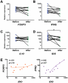
Figure 5. Relative expression of IDO decreases in CDL patients after treatment and is correlated with IFNG.
RNA was isolated from biopsies of lesions from CDL patients (n = 11) before and after treatment and the expression of FOXP3 (A), IFNG (B), IL10 (C) and IDO (D) was measured by qRT-PCR. The expression of each gene relative to healthy skin of four normal controls was calculated using the ΔΔCT method. E. Correlation between the relative expression of IDO and FOXP3 (left panel) or IFNG (right panel). *p<0.05, Wilcoxon signed-rank test.
Figure 6
Figure 6. IL-10 does not mediate suppression of effector functions by CD4+CD25+ cells.
IL-10 was measured in supernatants from the CD4+CD25-CD4+CD25+ co-cultures by ELISA. The level of IL-10 in the absence (1∶0 ratio) or presence (1∶1 ratio) of CD4+CD25+ cells is shown for AI (n = 6), CDL patients before treatment (n = 14), CDL patients after treatment (n = 11) and all co-cultures combined (n = 31). **p<0.01, Wilcoxon signed-rank test. Means with SEM are shown.

Similar articles

Cited by

References

    1. Murray HW, Berman JD, Davies CR, Saravia NG. Advances in leishmaniasis. Lancet. 2005;366:1561–1577. - PubMed
    1. Saravia NG, Valderrama L, Labrada M, Holguín AF, Navas C, et al. The relationship of Leishmania braziliensis subspecies and immune response to disease expression in New World leishmaniasis. J Infect Dis. 1989;159:725–735. - PubMed
    1. Carvalho EM, Johnson WD, Barreto E, Marsden PD, Costa JL, et al. Cell mediated immunity in American cutaneous and mucosal leishmaniasis. J Immunol. 1985;135:4144–4148. - PubMed
    1. Castilho TM, Goldsmith-Pestana K, Lozano C, Valderrama L, Saravia NG, et al. Murine model of chronic L. (Viannia) panamensis infection: role of IL-13 in disease. Eur J Immunol. 2010;40:2816–2829. - PMC - PubMed
    1. Díaz YR, Rojas R, Valderrama L, Saravia NG. T-bet, GATA-3, and Foxp3 expression and Th1/Th2 cytokine production in the clinical outcome of human infection with Leishmania (Viannia) species. J Infect Dis. 2010;202:406–415. - PMC - PubMed

Publication types

MeSH terms

Substances