Oncogenic splicing factor SRSF1 is a critical transcriptional target of MYC
- PMID: 22545246
- PMCID: PMC3334311
- DOI: 10.1016/j.celrep.2011.12.001
Oncogenic splicing factor SRSF1 is a critical transcriptional target of MYC
Abstract
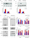
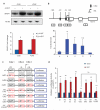
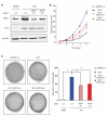
The SR protein splicing factor SRSF1 is a potent proto-oncogene that is frequently upregulated in cancer. Here, we show that SRSF1 is a direct target of the transcription factor oncoprotein MYC. These two oncogenes are significantly coexpressed in lung carcinomas, and MYC knockdown downregulates SRSF1 expression in lung-cancer cell lines. MYC directly activates transcription of SRSF1 through two noncanonical E-boxes in its promoter. The resulting increase in SRSF1 protein is sufficient to modulate alternative splicing of a subset of transcripts. In particular, MYC induction leads to SRSF1-mediated alternative splicing of the signaling kinase MKNK2 and the transcription factor TEAD1. SRSF1 knockdown reduces MYC's oncogenic activity, decreasing proliferation and anchorage-independent growth. These results suggest a mechanism for SRSF1 upregulation in tumors with elevated MYC and identify SRSF1 as a critical MYC target that contributes to its oncogenic potential by enabling MYC to regulate the expression of specific protein isoforms through alternative splicing.
Figures


Similar articles
-
Posttranscriptional Regulation of Splicing Factor SRSF1 and Its Role in Cancer Cell Biology.Biomed Res Int. 2015;2015:287048. doi: 10.1155/2015/287048. Epub 2015 Jul 26. Biomed Res Int. 2015. PMID: 26273603 Free PMC article. Review.
-
Alternative splicing of SLC39A14 in colorectal cancer is regulated by the Wnt pathway.Mol Cell Proteomics. 2011 Jan;10(1):M110.002998. doi: 10.1074/mcp.M110.002998. Epub 2010 Oct 11. Mol Cell Proteomics. 2011. PMID: 20938052 Free PMC article.
-
Hepatocyte growth factor enhances alternative splicing of the Kruppel-like factor 6 (KLF6) tumor suppressor to promote growth through SRSF1.Mol Cancer Res. 2012 Sep;10(9):1216-27. doi: 10.1158/1541-7786.MCR-12-0213. Epub 2012 Aug 2. Mol Cancer Res. 2012. PMID: 22859706
-
Phosphorylation of SRSF1 by SRPK1 regulates alternative splicing of tumor-related Rac1b in colorectal cells.RNA. 2014 Apr;20(4):474-82. doi: 10.1261/rna.041376.113. Epub 2014 Feb 18. RNA. 2014. PMID: 24550521 Free PMC article.
-
Emerging functions of SRSF1, splicing factor and oncoprotein, in RNA metabolism and cancer.Mol Cancer Res. 2014 Sep;12(9):1195-204. doi: 10.1158/1541-7786.MCR-14-0131. Epub 2014 May 7. Mol Cancer Res. 2014. PMID: 24807918 Free PMC article. Review.
Cited by
-
Mechanical stress during confined migration causes aberrant mitoses and c-MYC amplification.Proc Natl Acad Sci U S A. 2024 Jul 16;121(29):e2404551121. doi: 10.1073/pnas.2404551121. Epub 2024 Jul 11. Proc Natl Acad Sci U S A. 2024. PMID: 38990945 Free PMC article.
-
SRSF1 acts as an IFN-I-regulated cellular dependency factor decisively affecting HIV-1 post-integration steps.Front Immunol. 2022 Nov 15;13:935800. doi: 10.3389/fimmu.2022.935800. eCollection 2022. Front Immunol. 2022. PMID: 36458014 Free PMC article.
-
Genetic characterization of SF3B1 mutations in single chronic lymphocytic leukemia cells.Leukemia. 2013 Nov;27(11):2264-7. doi: 10.1038/leu.2013.155. Epub 2013 May 20. Leukemia. 2013. PMID: 23685408 Free PMC article. Clinical Trial. No abstract available.
-
Splicing-factor oncoprotein SRSF1 stabilizes p53 via RPL5 and induces cellular senescence.Mol Cell. 2013 Apr 11;50(1):56-66. doi: 10.1016/j.molcel.2013.02.001. Epub 2013 Mar 7. Mol Cell. 2013. PMID: 23478443 Free PMC article.
-
Posttranscriptional Regulation of Splicing Factor SRSF1 and Its Role in Cancer Cell Biology.Biomed Res Int. 2015;2015:287048. doi: 10.1155/2015/287048. Epub 2015 Jul 26. Biomed Res Int. 2015. PMID: 26273603 Free PMC article. Review.
References
-
- Adhikary S, Eilers M. Transcriptional regulation and transformation by Myc proteins. Nat. Rev. Mol. Cell Biol. 2005;6:635–645. - PubMed
-
- Amati B, Land H. Myc-Max: a transcription factor network controlling cell cycle progression, differentiation and death. Curr. Opin. Genet. Dev. 1994;4:102–108. - PubMed
-
- Biamonti G, Bassi MT, Cartegni L, Mechta F, Buvoli M, Cobianchi F, Riva S. Human hnRNP protein A1 gene expression. Structural and functional characterization of the promoter. J. Mol. Biol. 1993;230:77–89. - PubMed
Publication types
MeSH terms
Substances
Grants and funding
LinkOut - more resources
Full Text Sources
Other Literature Sources
Research Materials

