Review
. 2012 Nov;36(6):1046-82.
doi: 10.1111/j.1574-6976.2012.00342.x. Epub 2012 May 24.

Surface organelles assembled by secretion systems of Gram-negative bacteria: diversity in structure and function

Affiliations
Review

Surface organelles assembled by secretion systems of Gram-negative bacteria: diversity in structure and function

David G Thanassi et al. FEMS Microbiol Rev. 2012 Nov.

Abstract

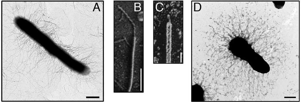
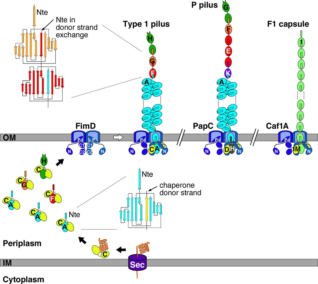
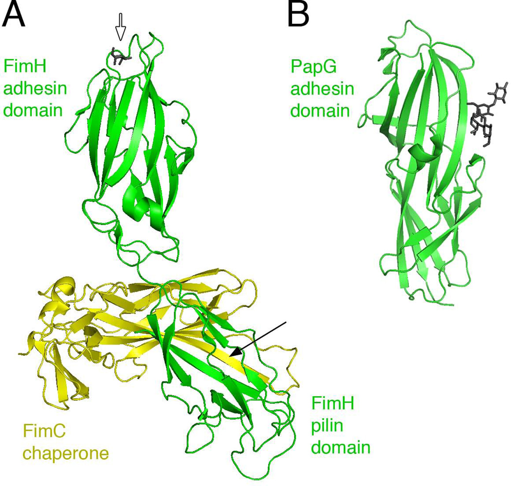
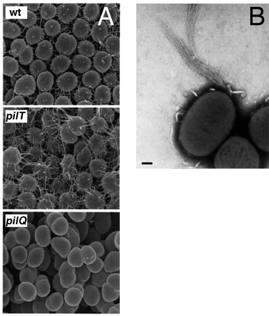
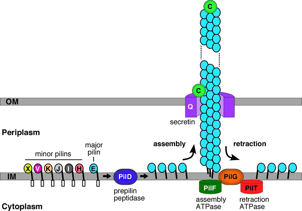
Gram-negative bacteria express a wide variety of organelles on their cell surface. These surface structures may be the end products of secretion systems, such as the hair-like fibers assembled by the chaperone/usher (CU) and type IV pilus pathways, which generally function in adhesion to surfaces and bacterial-bacterial and bacterial-host interactions. Alternatively, the surface organelles may be integral components of the secretion machinery itself, such as the needle complex and pilus extensions formed by the type III and type IV secretion systems, which function in the delivery of bacterial effectors inside host cells. Bacterial surface structures perform functions critical for pathogenesis and have evolved to withstand forces exerted by the external environment and cope with defenses mounted by the host immune system. Given their essential roles in pathogenesis and exposed nature, bacterial surface structures also make attractive targets for therapeutic intervention. This review will describe the structure and function of surface organelles assembled by four different Gram-negative bacterial secretion systems: the CU pathway, the type IV pilus pathway, and the type III and type IV secretion systems.


Figures

Fig. 1
Fig. 1
Electron micrographs of pili assembled by the CU pathway. (A) An E. coli bacterium expressing P pili. (B and C) High-resolution, freeze-etch images of individual E. coli P and type 1 pili, respectively, showing distal linear tip fibers and helical pilus rods. (D) A Y. pestis bacterium expressing F1 capsule. Scale bars = 500 nm (A), 100 nm (B), 20 nm (C), and 500 nm (D). Images in (A–D) reproduced with permission from (Kuehn et al., 1992; Jones et al., 1995; Runco et al., 2008; Li et al., 2010), respectively.
Fig. 2
Fig. 2
Model for pilus biogenesis by the CU pathway. The assembly steps for E. coli type 1 pili are shown, together with models for fully assembled E. coli type 1 and P pili, and the Y. pestis F1 capsule. The Fim, Pap, and Caf proteins are indicated by single letters (H, FimH; C, FimC; etc.). Pilus subunits enter the periplasm as unfolded polypeptides via the Sec pathway. The subunits fold upon interaction with the periplasmic chaperone, forming stable complexes via donor strand complementation. Assembly and secretion of the pilus fiber occurs at the OM usher, where chaperone-subunit interactions are replaced with subunit-subunit interactions via the donor strand exchange reaction. Topology diagrams are shown depicting the donor strand complementation and exchange reactions occurring in the periplasmic and pilus fiber, respectively. The dimeric ushers are depicted with the plug domain that gates the channel shut (P) and the periplasmic N and C domains indicated. The N domain forms the initial binding site for chaperone-subunit complexes and the C domains provide a second binding site for the assembling pilus fiber. Chaperone-adhesin complexes have highest affinity for the usher and initiate pilus assembly by binding to the usher N domain.
Fig. 3
Fig. 3
Structures of the FimH and PapG adhesins. (A) Crystal structure of the FimCH complex with bound mannose (PDB ID: 1KLF; (Hung et al., 2002)). The FimH adhesin and pilin domains are in green, with the bound mannose at the tip of the adhesin domain shown in dark gray stick representation (white arrow). The FimC chaperone is in yellow; the black arrow indicates the FimC β-strand engaged in donor strand complementation with the FimH pilin domain. (B) Crystal structure of the PapG adhesin domain with bound globoside (PDB ID: 1J8R; (Dodson et al., 2001)). The adhesin domain is in green and the tetrasaccharide bound at the side of the adhesin is in dark gray. Images were generated using PyMOL.
Fig. 4
Fig. 4
Electron micrographs of bacteria expressing T4P. (A) Scanning EM images showing T4P expressed by wild-type, and pilT and pilQ mutant N. gonorrhoeae. The pilT bacteria are hyperpiliated compared to wild-type, and the pilQ bacteria lack pili. (B) Micrograph of BFP expressed by EPEC showing a polar bundle of pili. Scale bar = 200 nm. Images in (A and B) reproduced with permission from (Wolfgang et al., 2000) and (Stone et al., 1996), respectively.
Fig. 5
Fig. 5
Model for biogenesis of T4P by N. meningitidis. The major (PilE) and minor (PilH, I, J, K, V and X) pilins are anchored in the inner membrane by their hydrophobic N-termini, with the conserved prepilin leader sequence exposed to the cytoplasm and globular head domain exposed to the periplasm. The leader sequence is cleaved and the mature pilins are N-methylated by the PilD prepilin peptidase. Processed pilin subunits are assembled into the pilus fiber from the periplasmic face of the inner membrane. PilF is the cytoplasmic ATPase that powers pilus assembly. The PilQ secretin provides the channel for secretion of the fiber across the outer membrane to the cell surface. Retraction of the pilus fiber is powered by the PilT cytoplasmic ATPase. During retraction, pilins are disassembled from the base of the pilus fiber and enter back into the inner membrane. The PilC protein functions as a tip-located adhesin, and also localizes to the outer membrane where it regulates pilus retraction.
Fig. 6
Fig. 6
Structures of the N. gonorrhoeae pilin and assembled T4P fiber. (A) Crystal structure of PilE (PDB ID: 2HI2; (Craig et al., 2006)). The N-terminal α-helix is shown in dark gray, the αβ-region in green, and the D-region in magenta with the conserved disulfide bond in yellow. The phosphoethanolamine and disaccharide modifications to serine-68 and -63, respectively, in the αβ-region are shown in stick representation. (B) Atomic model of the T4P fiber based on docking of the PilE structure into a cryoEM density map (PDB ID: 2HIL; (Craig et al., 2006)). The pilin subunits are shown in surface representation, with their N-terminal α-helices in dark gray and surface-exposed C-terminal globular domains colored according to the individual pilin subunits. The bottom-most pilin is depicted in ribbon representation in light green. Images were generated using PyMOL.
Fig. 7
Fig. 7
Electron micrographs of osmotically shocked S. enterica serovar Typhimurium bacteria. (Left) Nonflagellated ΔflhC S. Typhimurium exhibits needle complexes on the bacterial envelope. Note the depression at the insertion point of the needle complex (closed arrow). (Right) An S. Typhimurium fliK mutant exhibits flagellar polyhook basal bodies that span the inner and outer membranes. Scale bar = 100 nm. Reproduced with permission from (Kubori et al., 1998).
Fig. 8
Fig. 8
Structures of LcrV and the putative tip complex. (A) Ribbon diagram of LcrV (Derewenda et al., 2004) colored N (blue) to C (red) termini. (B) Overlay of the C-terminal helices of MxiH (red, residues 45–75) and LcrV (blue, residues 287–317), with all but the overlaid region made transparent to aid visualization. (C) Model of an LcrV tip complex (surface representation, gray) docked onto the tip of a T3SS needle. Reprinted with permission from (Deane et al., 2006). (D) Locations of protective epitopes in LcrV. Ribbon diagram of LcrV structure (PDB ID: 1R6F; (Derewenda et al., 2004)) shown in the same orientation as in panel A. Conformational epitope recognized by protective monoclonal antibody 7.3 (amino acids 135–275) (Hill et al., 1997; Hill et al., 2009) is shown in dark blue. Linear epitope recognized by protective monoclonal antibody BA5 (residues 196–225) (Eisele & Anderson, 2009; Quenee et al., 2010) is shown in light blue. Epitopes recognized by non-protective AH1 monoclonal antibody (Eisele & Anderson, 2009), corresponding to amino acids 76–105 of LcrV (D. Anderson, personal communication), are shown in magenta. Image was generated using PyMOL.
Fig. 9
Fig. 9
STEM images of negatively stained T3SS needles. (A) Characteristic tip complexes (arrow), comprising a head, a neck, and a base, of wild-type needles isolated from Y. enterocolitica grown in secretion-permissive conditions. (B) Needles formed by lcrV mutant bacteria. The needles of lcrV mutant bacteria are distinctly pointed at one end (asterisk). Scale bar = 20 nm. Reprinted with permission from (Mueller et al., 2005).
Fig. 10
Fig. 10
T4SS structures and mechanisms of action of the T4SS ATPases in substrate transfer and machine biogenesis. (A) Upper: CryoEM (left) and X-ray (right) structures of the VirB7/VirB9/VirB10 core complex from pKM101. Lower: X-ray structure of the VirD4-like TrwB hexamer from plasmid R388; the N-terminal TMD was modeled onto the X-ray structure of the TrwB soluble domain. X-ray structure of B. suis VirB11 hexamer. Model of VirB4 dimer (left, schematic of predicted VirB4 topology; right, SAXS model of a L. pneumophila VirB4 monomer superimposed with the VirB4 C-terminus from A. tumefaciens atomic model in ribbon representation). (B) Model depicting substrate docking with the VirD4 T4CP, delivery to VirB11, passage through a channel composed of VirB6 and VirB8 with a contribution by VirB4, and a VirB2 conduit. (C) Model depicting the contributions of VirB4 and VirB11 to dislocation of VirB2 pilin from the inner membrane to build the conjugative pilus. Red arrows denote the substrate translocation route (B) and VirB2 pilin dislocation and polymerization (C). Structures are reprinted with permission from: TrwB (Gomis-Ruth et al., 2001); VirB11 (Yeo et al., 2000); VirB4 (Durand et al., 2011); VirB7/VirB10/VirB10 core complex CryoEM (Fronzes et al., 2009) and X-ray crystallography (Chandran et al., 2009); VIrB5 (Yeo et al., 2003).
Fig. 11
Fig. 11
Examples of T4SS subunit acquisitions of structural folds for modulation of target cell interactions. (A) Some VirB6-like subunits possess C-terminal extensions implicated in surface exposure, e.g. Rickettsia spp. VirB6 (not shown), or extension through the T4SS channel and the recipient cell to the inner membrane where contact is established with the entry exclusion protein Eex, e.g. V. cholera SXT TraG and F plasmid TraG, which prevents redundant transfer to donor cells. (B) Several T4SSs possess variant forms of the VirB7/VirB9/VirB10 core complex subunits. X. citri VirB7XAC2622, a VirB7-like lipoprotein with an N-terminal (red) domain, implicated in binding VirB9XAC2620, and a C-terminal N0 domain (globular domain in yellow), which could mediate binding to target cells or substrate translocation through the T4SS. H. pylori VirB7-like CagT and VirB10-like CagY possess repeat domains implicated in forming a surface-variable sheath structure. (C) H. pylori VirB5-like CagL has an RGD motif implicated in binding of the β-integrin receptor on the host cell. Bartonella and Rickettsia spp. code for multiple copies of VirB2-like pilins, TrwL and VirB2, respectively, implicated in forming surface-variable pilins for immunomodulation. The X. citri VirB7 X-ray structure is reprinted with permission from (Souza et al., 2011). The modeled structure of VirB5-like CagL is reprinted with permission from (Backert et al., 2008). The schematics in Figs. B and C are reprinted with permission from (Alvarez-Martinez & Christie, 2009).

References

    1. Aas FE, Lovold C, Koomey M. An inhibitor of DNA binding and uptake events dictates the proficiency of genetic transformation in Neisseria gonorrhoeae: mechanism of action and links to Type IV pilus expression. Mol Microbiol. 2002;46:1441–1450. - PubMed
    1. Abraham SN, Sun D, Dale JB, Beachey EH. Conservation of the D-mannose-adhesion protein among type 1 fimbriated members of the family Enterobactericeae. Nature. 1988;336:682–684. - PubMed
    1. Akeda Y, Galan JE. Chaperone release and unfolding of substrates in type III secretion. Nature. 2005;437:911–915. - PubMed
    1. Akopyan K, Edgren T, Wang-Edgren H, Rosqvist R, Fahlgren A, Wolf-Watz H, Fallman M. Translocation of surface-localized effectors in type III secretion. Proc Natl Acad Sci U S A. 2011 - PMC - PubMed
    1. Alvarez-Martinez CE, Christie PJ. Biological diversity of prokaryotic type IV secretion systems. Microbiol Mol Biol Rev. 2009;73:775–808. - PMC - PubMed

Publication types

MeSH terms