. 2012 May;9(5):497-504.
doi: 10.3171/2012.1.PEDS11453.

Follow-up imaging to detect recurrence of surgically treated pediatric arteriovenous malformations

Affiliations

Follow-up imaging to detect recurrence of surgically treated pediatric arteriovenous malformations

Shih-Shan Lang et al. J Neurosurg Pediatr. 2012 May.

Abstract

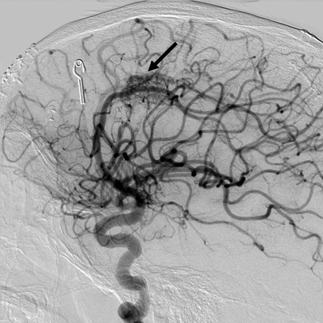
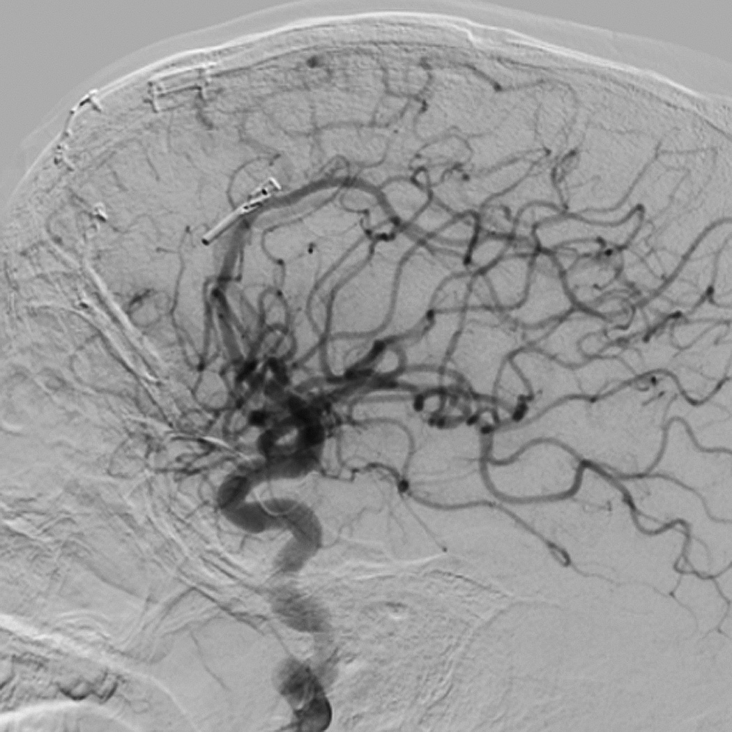
Object: The true postoperative incidence of arteriovenous malformation (AVM) recurrence in the pediatric population remains largely unreported. Some literature suggests that delayed imaging studies should be obtained at 6 months to 1 year after negative findings on a postoperative angiogram. The aim of this study was to describe the timing of AVM recurrences after resection and the neuroimaging modalities on which the recurrences were detected.

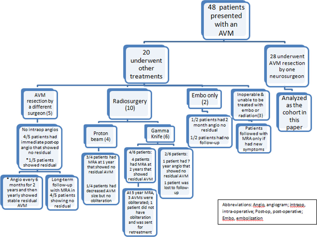
Methods: This study was performed in a retrospective cohort of all pediatric patients treated surgically for AVM resection by a single neurosurgeon between 2005 and 2010. Patients were followed after resection with MR angiography (MRA) or conventional angiography, when possible, at various time points. A visual scale for compactness of the initial AVM nidus was used, and the score was correlated with probability of recurrence after surgery.

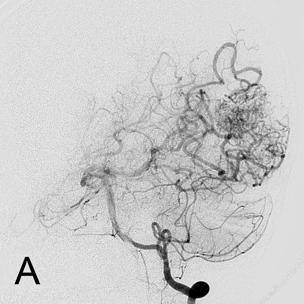
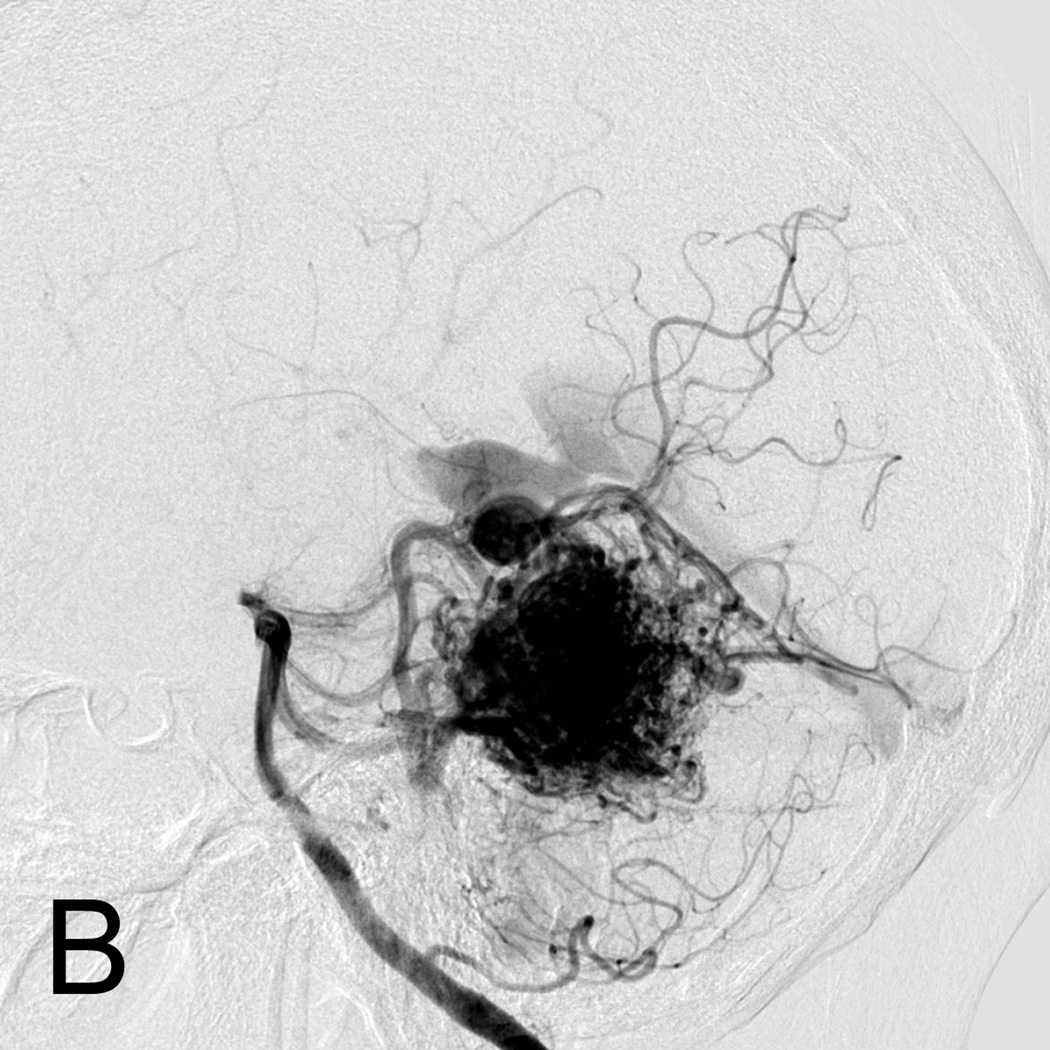
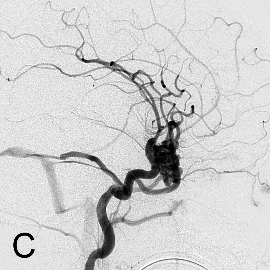
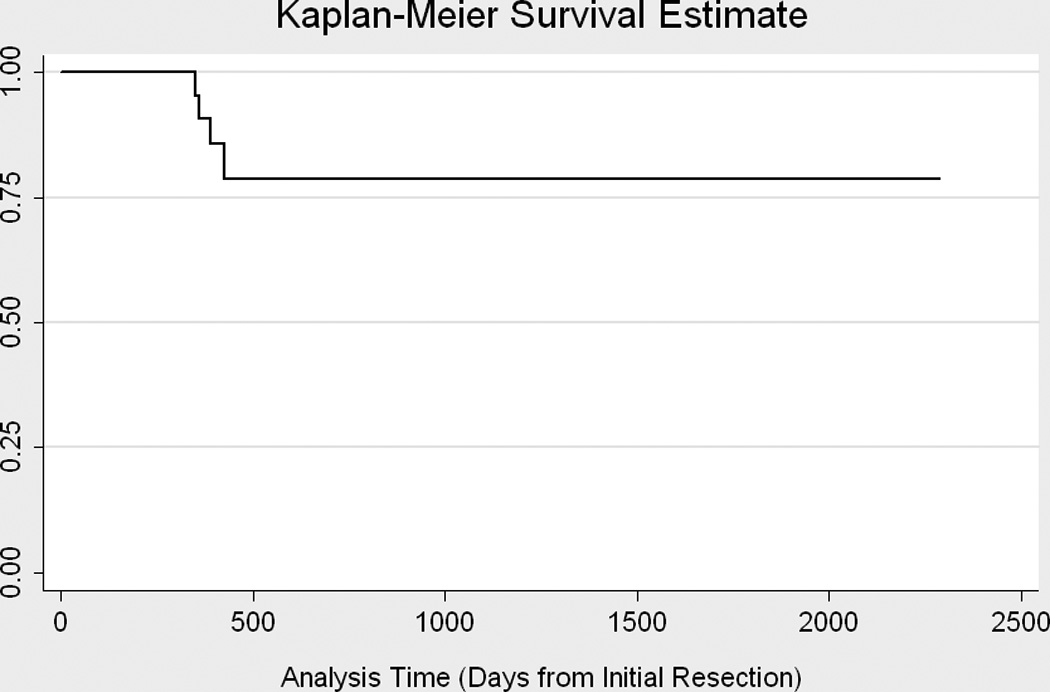
Results: A total of 28 patients (13 female, 15 male) underwent an AVM resection. In 18 patients (64.3%) an intraoperative angiogram was obtained. In 4 cases the intraoperative angiogram revealed residual AVM, and repeat resections were performed immediately. Recurrent AVMs were found in 4 children (14.3%) at 50, 51, 56, and 60 weeks after the initial resection. Recurrence risk was 0.08 per person-year. No patient with normal results on an angiogram obtained at 1 year developed a recurrence on either a 5-year angiogram or one obtained at 18 years of age. All patients with recurrence had a compactness score of 1 (diffuse AVM); a lower compactness score was associated with recurrence (p = 0.0003).

Conclusions: All recurrences in this cohort occurred less than 15 months from the initial resection. The authors recommend intraoperative angiography to help ensure complete resection at the time of the surgery. Follow-up vascular imaging is crucial for detecting recurrent AVMs, and conventional angiography is preferred because MRA can miss smaller AVMs. One-year follow-up imaging detected these recurrences, and no one who had negative results on an angiogram obtained at 1 year had a late recurrence. However, not all of the patients have been followed for 5 years or until 18 years of age, so longer follow-up is required for these patients. A lower compactness score predicted recurrent AVM in this cohort.


Conflict of interest statement

None of the authors has any disclosures or conflicts of interest.

Figures

Figure 1
Figure 1
Flow-diagram of 48 subjects presenting to our institution from 2005–2010.
Figure 2
Figure 2
Visual AVM nidus Compactness score A. AVM with compactness score of 1 showing a diffuse nidus. B. AVM with compactness score of 2. C. Highly compact AVM nidus with compactness score of 3.
Figure 2
Figure 2
Visual AVM nidus Compactness score A. AVM with compactness score of 1 showing a diffuse nidus. B. AVM with compactness score of 2. C. Highly compact AVM nidus with compactness score of 3.
Figure 2
Figure 2
Visual AVM nidus Compactness score A. AVM with compactness score of 1 showing a diffuse nidus. B. AVM with compactness score of 2. C. Highly compact AVM nidus with compactness score of 3.
Figure 3
Figure 3
Time to Recurrence. Kaplain-Meyer Survival Curve.
Figure 4
Figure 4
Eight year old male who presented with papilledema during routine eye examination. (Patient 2, Table 4 A. T2-weighted axial MRI demonstrates serpiginous flow voids of various sizes in the left frontal lobe suggestive of AVM. B. Lateral left internal carotid artery injection digital subtraction angiogram demonstrates a 6 cm left frontal AVM fed predominantly by both anterior cerebral arteries (ACAs), but predominately by the left ACA with superficial drainage. A compactness score of 1 was assigned. C. Intra-operative angiography following resection was performed showing complete resection of the AVM on lateral view. D. Lateral left internal carotid artery injection digital subtraction angiogram 1 year post-resection demonstrating recurrence of the AVM (arrow). Patient underwent craniotomy for recurrent AVM resection. E. Lateral left internal carotid artery injection digital subtraction angiogram 1 year post-operatively after second resection demonstrating no recurrent AVM.
Figure 4
Figure 4
Eight year old male who presented with papilledema during routine eye examination. (Patient 2, Table 4 A. T2-weighted axial MRI demonstrates serpiginous flow voids of various sizes in the left frontal lobe suggestive of AVM. B. Lateral left internal carotid artery injection digital subtraction angiogram demonstrates a 6 cm left frontal AVM fed predominantly by both anterior cerebral arteries (ACAs), but predominately by the left ACA with superficial drainage. A compactness score of 1 was assigned. C. Intra-operative angiography following resection was performed showing complete resection of the AVM on lateral view. D. Lateral left internal carotid artery injection digital subtraction angiogram 1 year post-resection demonstrating recurrence of the AVM (arrow). Patient underwent craniotomy for recurrent AVM resection. E. Lateral left internal carotid artery injection digital subtraction angiogram 1 year post-operatively after second resection demonstrating no recurrent AVM.

References

    1. Ali MJ, Bendok BR, Rosenblatt S, Rose JE, Getch CC, Batjer HH. Recurrence of pediatric cerebral arteriovenous malformations after angiographically documented resection. Pediatr Neurosurg. 2003;39:32–38. - PubMed
    1. Andaluz N, Myseros JS, Sathi S, Crone KR, Tew JM., Jr Recurrence of cerebral arteriovenous malformations in children: report of two cases and review of the literature. Surg Neurol. 2004;62:324–330. discussion 330-321. - PubMed
    1. Bristol RE, Albuquerque FC, Spetzler RF, Rekate HL, McDougall CG, Zabramski JM. Surgical management of arteriovenous malformations in children. J Neurosurg. 2006;105:88–93. - PubMed
    1. Deruty R, Pelissou-Guyotat I, Amat D, Mottolese C, Bascoulergue Y, Turjman F, Gerard JP. Multidisciplinary treatment of cerebral arteriovenous malformations. Neurol Res. 1995;17:169–177. - PubMed
    1. Di Rocco C, Tamburrini G, Rollo M. Cerebral arteriovenous malformations in children. Acta Neurochir (Wien) 2000;142:145–156. discussion 156-148. - PubMed

Publication types

MeSH terms

LinkOut - more resources