. 2012 Jul;18(13-14):1520-8.
doi: 10.1089/ten.TEA.2011.0127. Epub 2012 Jun 7.

Bioceramic-mediated trophic factor secretion by mesenchymal stem cells enhances in vitro endothelial cell persistence and in vivo angiogenesis

Affiliations

Bioceramic-mediated trophic factor secretion by mesenchymal stem cells enhances in vitro endothelial cell persistence and in vivo angiogenesis

Jiawei He et al. Tissue Eng Part A. 2012 Jul.

Abstract

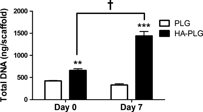
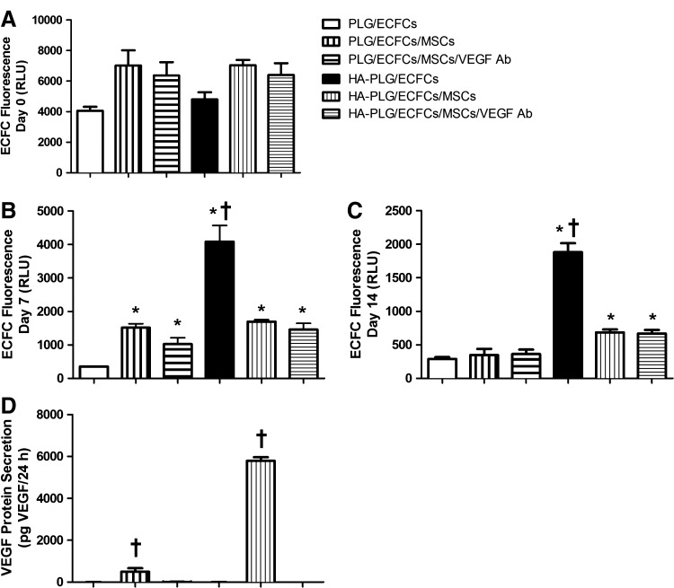
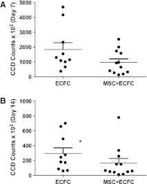
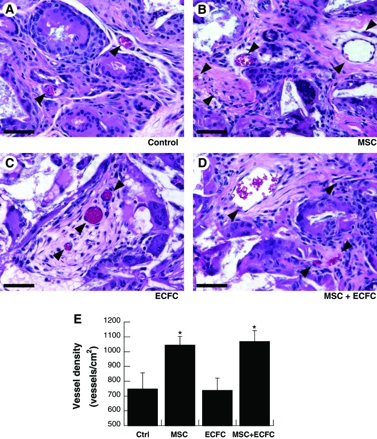
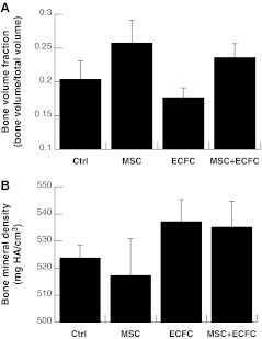
Mesenchymal stem cells (MSCs) seeded in composite implants formed of hydroxyapatite (HA) and poly (lactide-co-glycolide) (PLG) exhibit increased osteogenesis and enhanced angiogenic potential. Endothelial colony-forming cells (ECFCs) can participate in de novo vessel formation when implanted in vivo. The aim of this study was to determine the capacity of HA-PLG composites to cotransplant MSCs and ECFCs, with the goal of accelerating vascularization and resultant bone formation. The incorporation of HA into PLG scaffolds improved the efficiency of cell seeding and ECFC survival in vitro. We observed increases in mRNA expression and secretion of potent angiogenic factors by MSCs when cultured on HA-PLG scaffolds compared to PLG controls. Upon implantation into an orthotopic calvarial defect, ECFC survival on composite scaffolds was not increased in the presence of MSCs, nor did the addition of ECFCs enhance vascularization beyond increases observed with MSCs alone. Microcomputed tomography (micro-CT) performed on explanted calvarial tissues after 12 weeks revealed no significant differences between treatment groups for bone volume fraction (BVF) or bone mineral density (BMD). Taken together, these results provide evidence that HA-containing composite scaffolds seeded with MSCs can enhance neovascularization, yet MSC-secreted trophic factors do not consistently increase the persistence of co-transplanted ECFCs.


Figures

FIG. 1.
FIG. 1.
DNA quantification of MSC-seeded scaffolds was performed at days 0 and 7 on PLG and HA–PLG scaffolds. Values are mean±SEM (n=4); **p<0.01 versus PLG at day 0; ***p<0.001 versus PLG at day 7. p<0.001 versus HA–PLG at day 0. MSC, mesenchymal stem cell; PLG, poly (lactide-co-glycolide); HA, hydroxyapatite; SEM, standard error of the mean.
FIG. 2.
FIG. 2.
Quantitative polymerase chain reaction was used to analyze proangiogenic gene expression by MSCs on various scaffolds. (A) VEGFA, (B) PDGF, (C) FGF1, and (D) FGF2 expression were normalized to RPL13 expression and data were represented as ΔCt. Values are mean±SEM (n=4); *p<0.05 versus PLG at the same time point; **p<0.01 versus PLG at day 0; $p<0.05 versus PLG at day 0; ***p<0.001 versus expression at day 0.
FIG. 3.
FIG. 3.
(A–C) Fluorescent quantification of ECFCs seeded individually or in coculture on PLG and HA–PLG scaffolds at days 0, 7, and 14. (D) VEGF secretion by cells on scaffolds at day 7. Values are mean±SEM (n=4); *p<0.05 versus PLG, p<0.05 versus all groups. All groups except HA-PLG/ECFCs demonstrated significant reductions in fluorescence by day 7. ECFCs, endothelial colony-forming cells; VEGF, vascular endothelial growth factor; RLU, relative light units.
FIG. 4.
FIG. 4.
In vivo bioluminescent measurements used to measure luciferin-transduced ECFCs on scaffolds transplanted to cranial defects after (A) 7 and (B) 14 days. *p<0.05 versus MSC+ECFC.
FIG. 5.
FIG. 5.
Neovascularization of composite scaffolds after implantation for 4 weeks. Representative hematoxylin and eosin sections near the center of explanted scaffolds imaged at 40×(A–D). (Arrowheads denote vessels; scale bar=50 μm). (E) Vessel density within repair tissue; values are mean±SEM (n=5); *p<0.05 versus acellular control scaffolds. Color images available online at www.liebertpub.com/tea
FIG. 6.
FIG. 6.
micro-CT was used to quantify (A) BVF and (B) BMD after removal of calvarial tissue. No significant differences were detected. Micro-CT, microcomputed tomography; BVF, bone volume fraction; BMD, bone mineral density.

References

    1. Patel Z.S. Young S. Tabata Y. Jansen J.A. Wong M.E. Mikos A.G. Dual delivery of an angiogenic and an osteogenic growth factor for bone regeneration in a critical size defect model. Bone. 2008;43:931. - PMC - PubMed
    1. Kaigler D. Krebsbach P.H. Wang Z. West E.R. Horger K. Mooney D.J. Transplanted endothelial cells enhance orthotopic bone regeneration. J Dent Res. 2006;85:633. - PubMed
    1. Kumar S. Wan C. Ramaswamy G. Clemens T.L. Ponnazhagan S. Mesenchymal stem cells expressing osteogenic and angiogenic factors synergistically enhance bone formation in a mouse model of segmental bone defect. Mol Ther. 2010;18:1026. - PMC - PubMed
    1. Peng H. Usas A. Olshanski A. Ho A.M. Gearhart B. Cooper G.M., et al. VEGF improves, whereas sFlt1 inhibits, BMP2-induced bone formation and bone healing through modulation of angiogenesis. J Bone Miner Res. 2005;20:2017. - PubMed
    1. Leu A. Stieger S.M. Dayton P. Ferrara K.W. Leach J.K. Angiogenic response to bioactive glass promotes bone healing in an irradiated calvarial defect. Tissue Eng Part A. 2009;15:877. - PMC - PubMed

Publication types

MeSH terms

LinkOut - more resources