Review
. 2013 Oct 22:251:90-107.
doi: 10.1016/j.neuroscience.2012.04.044. Epub 2012 Apr 27.

Dendritic spine pathology in schizophrenia

Affiliations
Review

Dendritic spine pathology in schizophrenia

J R Glausier et al. Neuroscience. .

Abstract

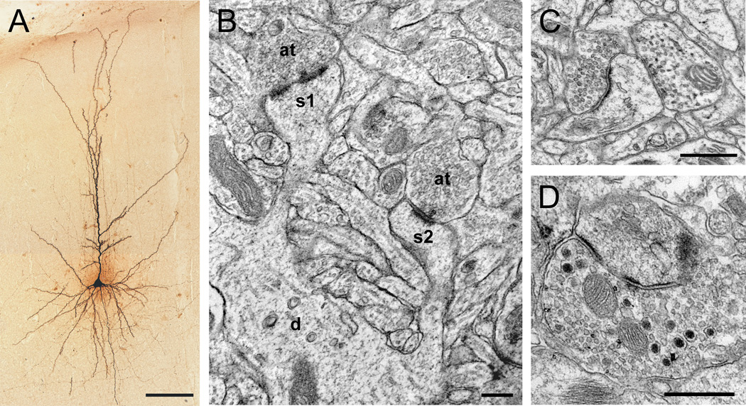
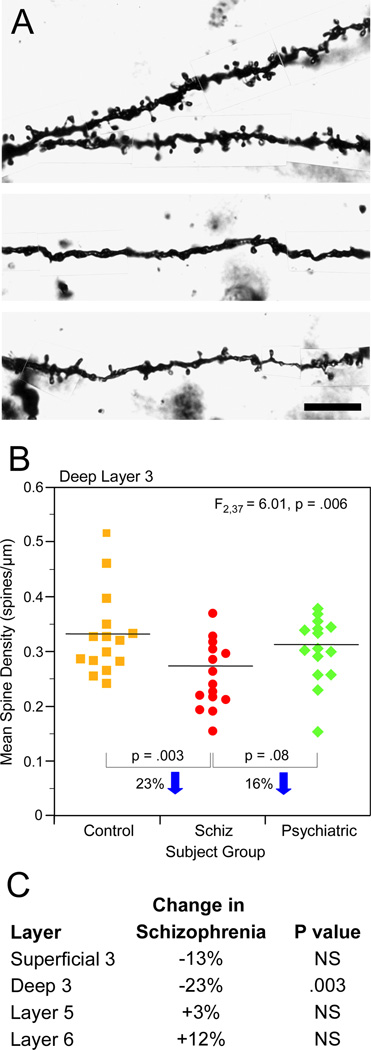
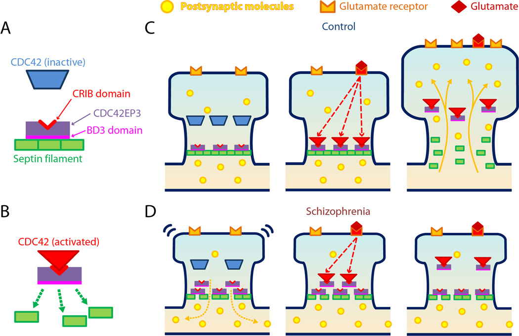
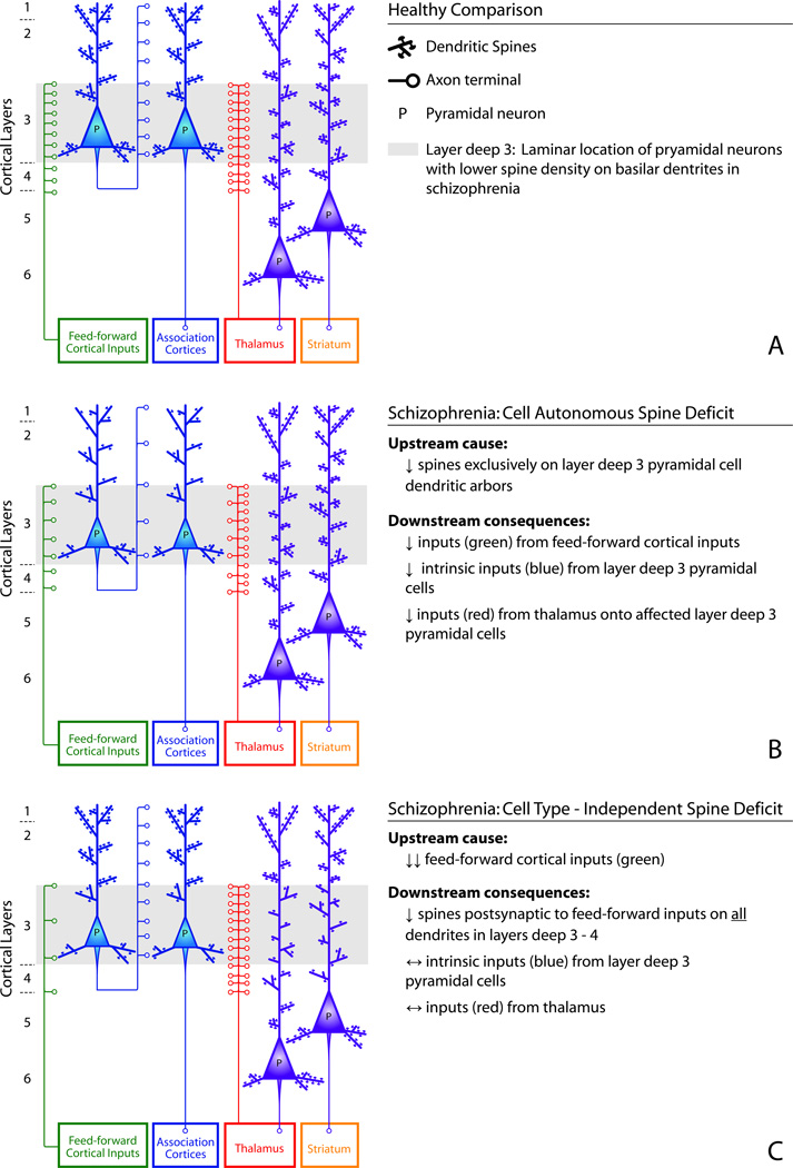
Schizophrenia is a neurodevelopmental disorder whose clinical features include impairments in perception, cognition and motivation. These impairments reflect alterations in neuronal circuitry within and across multiple brain regions that are due, at least in part, to deficits in dendritic spines, the site of most excitatory synaptic connections. Dendritic spine alterations have been identified in multiple brain regions in schizophrenia, but are best characterized in layer 3 of the neocortex, where pyramidal cell spine density is lower. These spine deficits appear to arise during development, and thus are likely the result of disturbances in the molecular mechanisms that underlie spine formation, pruning, and/or maintenance. Each of these mechanisms may provide insight into novel therapeutic targets for preventing or repairing the alterations in neural circuitry that mediate the debilitating symptoms of schizophrenia.

Keywords: 22q11 DS; 22q11 deletion syndrome; APDs; DISC1; DLPFC; GEFs; MD; MFT; MRI; MSN; PFC; PMI; SP-IR; TDL; VTA; antipsychotic drugs; development; disrupted in schizophrenia 1; dorsolateral prefrontal cortex; guanine exchange factors; laminar; magnetic resonance imaging; mediodorsal; medium spiny neuron; medium spiny neurons; mossy fiber terminations; postmortem intervals; prefrontal cortex; pruning; pyramidal cell; spinogenesis; spinophilin-immunoreactive; total dendritic length; ventral tegmental area.


Figures

Figure 1
Figure 1
Micrographs of pyramidal cell dendritic spines in the primate prefrontal cortex. A) Light micrograph of a biocytin-filled pyramidal cell showing the cell body, and basilar and apical dendritic shafts. B) Electron micrograph showing two spines (s1, s2) protruding from a dendritic shaft (d). Spine s1 is receiving a perforated asymmetric (presumably glutamatergic) synapse. C) Dendritic spine receiving an asymmetric (presumably glutamatergic) synapse and a symmetric (inhibitory or catecholaminergic) synapse. Note the difference in the appearance of the synaptic vesicles in each presynaptic axon terminal. D) Axon terminal containing small, round vesicles characteristic of glutamate, and large, dense-core vesicles characteristic of neuropeptides, forming an asymmetric synapse onto a spine. Scale bar is 100 µm in A, 200 nm in B, and 500 nm in C–D.
Figure 2
Figure 2
Decreased layer 3 spine density in the dorsolateral prefrontal cortex (DLPFC) of schizophrenia subjects. A) Golgi-impregnated basilar dendrites and spines from a healthy comparison subject (top) and two schizophrenia subjects (bottom). Spine density is qualitatively decreased in the schizophrenia subjects relative to the healthy comparison subject. B) Scatter plot demonstrating lower layer deep 3 spine density in schizophrenia subjects relative to healthy comparison and psychiatrically ill subjects. C) Within the same subjects, lower DLPFC spine density is specific to layer deep 3. Scale bar is 10 µm in A. Adapted from Lewis and Gonzalez-Burgos (2008).
Figure 3
Figure 3
Schematic diagram of Cdc42-Cdc42EP3-septin interactions and their proposed roles in spine dysfunction in schizophrenia. A) Cdc42EP3 binds to septins via a BD3 domain, inducing the assembly of septin filaments. Inactive Cdc42 cannot bind to Cdc42EP3. B) Once activated, Cdc42 can bind to and inhibit Cdc42EP3 via its CRIB domain, disrupting septin filament formation. C) In the normal state, Cdc42EP3 consolidates septin filaments in the spine neck, effectively forming a barrier for molecular diffusion with the parent dendrite (left). Transient activation of Cdc42 by glutamate stimulation inhibits the Cdc42EP3-mediated assembly of the septin filament barrier (middle), enabling molecular diffusion with the parent dendrite (right). C) In schizophrenia, decreased expression of septin 7 mRNA contributes to impaired septin filament formation at the spine neck, limiting the retention of postsynaptic molecules that are critical for spine structure and function in the spine head (left). Furthermore, lower levels of Cdc42 and increased levels of Cdc42EP3 lead to a reduced capacity for glutamatergic signaling to open the septin barrier (middle), limiting synaptic plasticity and possibly contributing to spine loss (right). Cell division cycle 42 (Cdc42), Cdc42 effector protein 3(Cdc42EP3). Adapted from Ide and Lewis 2010.
Figure 4
Figure 4
Two alternative models of potential cause and consequences of layer deep 3 spine deficits in schizophrenia.

References

    1. Abi-Dargham A, Mawlawi O, Lombardo I, Gill R, Martinez D, Huang Y, Hwang DR, Keilp J, Kochan L, van Heertum R, Gorman JM, Laruelle M. Prefrontal dopamine D1 receptors and working memory in schizophrenia. J Neurosci. 2002;22:3708–3719. - PMC - PubMed
    1. Abi-Dargham A, Xu X, Thompson JL, Gil R, Kegeles LS, Urban NB, Narendran R, Hwang DR, Laruelle M, Slifstein M. Increased prefrontal cortical D1 receptors in drug naive patients with schizophrenia: a PET study with [11C]NNC112. J Psychopharmacol. 2011 [epub ahead of print]. - PubMed
    1. Ahmed B, Anderson JC, Douglas RJ, Martin KAC, Nelson JC. Polyneuronal innervation of spiny stellate neurons in cat visual cortex. J Comp Neurol. 1994;341:39–49. - PubMed
    1. Akil M, Pierri JN, Whitehead RE, Edgar CL, Mohila C, Sampson AR, Lewis DA. Lamina-specific alterations in the dopamine innervation of the prefrontal cortex in schizophrenic subjects. Am J Psychiatry. 1999;156:1580–1589. - PubMed
    1. Aleman A, Kahn RS, Selten JP. Sex differences in the risk of schizophrenia: evidence from meta-analysis. Arch Gen Psychiatry. 2003;60:565–571. - PubMed

Publication types

MeSH terms