

IgG4-related inflammatory pseudotumor of the central nervous system responsive to mycophenolate mofetil
- PMID: 22546342
- PMCID: PMC3366053
- DOI: 10.1016/j.jns.2012.04.010
IgG4-related inflammatory pseudotumor of the central nervous system responsive to mycophenolate mofetil
Abstract
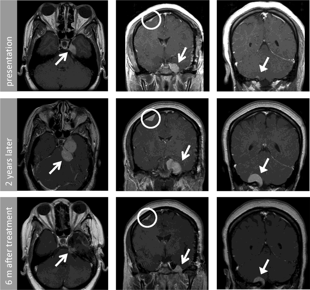
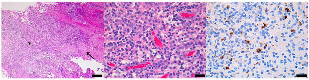
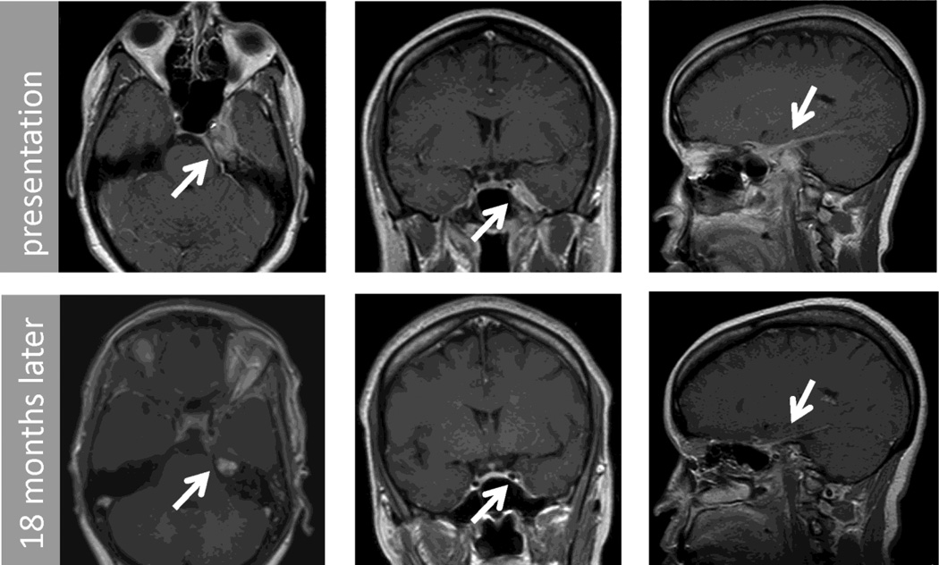
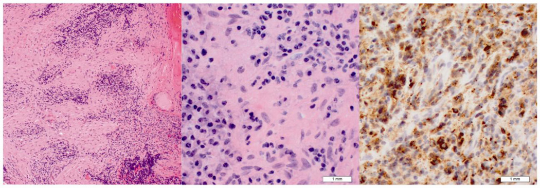
Orbital apex and skull base masses often present with neuro-ophthalmic signs and symptoms. Though the localization of these syndromes and visualization of the responsible lesion on imaging is typically straightforward, definitive diagnosis usually relies on biopsy. Immunohistochemistry is important for categorization and treatment planning. IgG4-related disease is emerging as a pathologically defined inflammatory process that can occur in multiple organ systems. We present two patients with extensive inflammatory mass lesions of the central nervous system with immunohistochemistry positive for IgG4 and negative for ALK-1 as examples of meningeal based IgG4-related inflammatory pseudotumors. In both patients, there was treatment response to mycophenolate mofetil.
Copyright © 2012 Elsevier B.V. All rights reserved.
Conflict of interest statement
The authors disclose no financial or personal relationships with other people or organizations that could inappropriately influence this work
Figures
References
-
- Swain RS, Tihan T, Horvai AE, Di Vizio D, Loda M, Burger PC, et al. Inflammatory myofibroblastic tumor of the central nervous system and its relationship to inflammatory pseudotumor. Human pathology. 2008;39(3):410–419. - PubMed
-
- Bateman AC, Deheragoda MG. IgG4 related systemic sclerosing diseases: an emerging and under diagnosed condition. Histopathology. 2009;55(4):373–383. - PubMed
-
- Lui PCW, Fan YS, Wong SS, Chan ANH, Wong G, Chau TKF, et al. Inflammatory pseudotumors of the central nervous system. Human pathology. 2009;40(11):1611–1617. - PubMed
-
- Yamamoto H, Yamaguchi H, Aishima S, Oda Y, Kohashi K, Oshiro Y, et al. Inflammatory myofibroblastic tumor versus IgG4-related sclerosing disease and inflammatory pseudotumor: a comparative clinicopathologic study. The American journal of surgical pathology. 2009;33(9):1330. - PubMed
-
- Hausler M, Schaade L, Ramaekers VT, Doenges M, Heimann G, Sellhaus B. Inflammatory pseudotumors of the central nervous system: report of 3 cases and a literature review. Human pathology. 2003;34(3):253–262. - PubMed
Publication types
MeSH terms
Substances
Grants and funding
LinkOut - more resources
Full Text Sources
Medical
Miscellaneous
