. 2012 Apr 30;4(4):33.
doi: 10.1186/gm332.

Cancer detection and biopsy classification using concurrent histopathological and metabolomic analysis of core biopsies

Affiliations

Cancer detection and biopsy classification using concurrent histopathological and metabolomic analysis of core biopsies

Meredith V Brown et al. Genome Med. .

Abstract

Background: Metabolomics, the non-targeted interrogation of small molecules in a biological sample, is an ideal technology for identifying diagnostic biomarkers. Current tissue extraction protocols involve sample destruction, precluding additional uses of the tissue. This is particularly problematic for high value samples with limited availability, such as clinical tumor biopsies that require structural preservation to histologically diagnose and gauge cancer aggressiveness. To overcome this limitation and increase the amount of information obtained from patient biopsies, we developed and characterized a workflow to perform metabolomic analysis and histological evaluation on the same biopsy sample.

Methods: Biopsies of ten human tissues (muscle, adrenal gland, colon, lung, pancreas, small intestine, spleen, stomach, prostate, kidney) were placed directly in a methanol solution to recover metabolites, precipitate proteins, and fix tissue. Following incubation, biopsies were removed from the solution and processed for histology. Kidney and prostate cancer tumor and benign biopsies were stained with hemotoxylin and eosin and prostate biopsies were subjected to PIN-4 immunohistochemistry. The methanolic extracts were analyzed for metabolites on GC/MS and LC/MS platforms. Raw mass spectrometry data files were automatically extracted using an informatics system that includes peak identification and metabolite identification software.

Results: Metabolites across all major biochemical classes (amino acids, peptides, carbohydrates, lipids, nucleotides, cofactors, xenobiotics) were measured. The number (ranging from 260 in prostate to 340 in colon) and identity of metabolites were comparable to results obtained with the current method requiring 30 mg ground tissue. Comparing relative levels of metabolites, cancer tumor from benign kidney and prostate biopsies could be distinguished. Successful histopathological analysis of biopsies by chemical staining (hematoxylin, eosin) and antibody binding (PIN-4, in prostate) showed cellular architecture and immunoreactivity were retained.

Conclusions: Concurrent metabolite extraction and histological analysis of intact biopsies is amenable to the clinical workflow. Methanol fixation effectively preserves a wide range of tissues and is compatible with chemical staining and immunohistochemistry. The method offers an opportunity to augment histopathological diagnosis and tumor classification with quantitative measures of biochemicals in the same tissue sample. Since certain biochemicals have been shown to correlate with disease aggressiveness, this method should prove valuable as an adjunct to differentiate cancer aggressiveness.


Figures

Figure 1
Figure 1
Schematic outline of method workflow. Flow diagram of the intact biopsy extraction protocol and tissue grinding protocol for a tissue biopsy and a 30 mg tissue piece. RT, room temperature.
Figure 2
Figure 2
Number and identity of metabolites obtained with intact biopsy method and the standard ground tissue extraction method. The total number of metabolites detected using each sampling and extraction protocol (30 mg tissue, intact biopsy, ground biopsy) is shown in the rectangles at the bottom of the figure. The Venn diagram represents the overlap in the identity of metabolites detected using each method. The vast majority (266) of metabolites are detected using all three methods. Metabolites were extracted from intact biopsies with 80% methanol.
Figure 3
Figure 3
Histochemical staining of biopsy samples treated with methanol or ethanol as the extraction solvent. Human prostate biopsies from benign or cancer tumor tissue were processed using the intact biopsy method in either 80% methanol or 70% ethanol or fixed in formalin followed by paraffin embedding and sectioning. The resulting sections were then stained with hematoxylin and eosin.
Figure 4
Figure 4
Histology of prostate biopsy samples. Human prostate biopsy samples were processed using the intact biopsy method in 80% methanol followed by paraffin embedding and sectioning. (a) Prostate section processed for immunohistochemistry using PIN4 stain where red indicates racemase and brown indicates p63 and basal keratin. (b) An immediately adjacent section stained with hematoxylin and eosin (H & E). Black arrows indicate prostatic adenocarcinoma and blue arrows indicate benign glands.
Figure 5
Figure 5
Representative histology images from kidney biopsies show tissue structure is retained. Patient-matched (a) benign and (b) cancer tumor kidney biopsies were processed using the intact biopsy workflow and stained with hematoxylin and eosin. Scale bars, 50 μm.
Figure 6
Figure 6
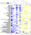
Cancer tumor and benign kidney samples can be separated using hierarchical cluster analysis. The 69 metabolites identified as significantly different (P ≤ 0.05) between cancer tumor and matched benign kidney tissue from six patients were used to generate the cluster based on Euclidean distance. Cancer tumor or benign samples were determined by histopathology evaluation. Metabolites are listed on the y-axis. Each patient is represented by a number (1 to 6) on the x-axis. Cancer tumor (C) and matched benign (B) samples were used for the analysis. Four of six cancer tumor samples were assigned to the same major cluster and five of six benign samples were assigned to the same major cluster.
Figure 7
Figure 7
Principal components analysis of kidney biopsies to distinguish cancer tumor from benign biopsies. The metabolites identified as significant (P ≤ 0.05) between cancer tumor-containing and benign kidney biopsies by matched pairs t-test were used to construct the principal components analysis. Blue, cancer tumor samples; yellow, benign samples. The six nephrectomy patients are each indicated by a shape: circle, patient 1; square, patient 2; upward triangle, patient 3; downward triangle, patient 4; left pointing triangle, patient 5; right pointing triangle, patient 6.
Figure 8
Figure 8
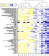
Hierarchical cluster analysis of cancer tumor and benign prostate samples. The 83 metabolites determined to be significantly different (P < 0.05) between cancer tumor and matched benign tissue from eight patients were used to generate the cluster based on Euclidean distance. Metabolites are listed on the y-axis. Each patient is represented by a number (1 to 8) on the x-axis. Histologically determined cancer tumor (C) and matched benign (B) samples were used for the analysis. Cancer tumor and benign biopsies fall into two major clusters. Seven of eight cancer tumor and seven of eight benign samples clustered as predicted by the histological analysis of the biopsy.

References

    1. Iczkowski KA, Lucia MS. Current perspectives on Gleason grading of prostate cancer. Curr Urol Rep. 2011;12:216–222. doi: 10.1007/s11934-011-0181-5. - DOI - PubMed
    1. Netto GJ, Eisenberger M, Epstein JI. Interobserver variability in histologic evaluation of radical prostatectomy between central and local pathologists: findings of TAX 3501 multinational clinical trial. Urology. 2011;77:1155–1160. doi: 10.1016/j.urology.2010.08.031. - DOI - PMC - PubMed
    1. Ficarra V, Martignoni G, Maffei N, Brunelli M, Novara G, Zanolla L, Pea M, Artibani W. Original and reviewed nuclear grading according to the Fuhrman system: a multivariate analysis of 388 patients with conventional renal cell carcinoma. Cancer. 2005;103:68–75. doi: 10.1002/cncr.20749. - DOI - PubMed
    1. May M, Brookman-Amissah S, Roigas J, Hartmann A, Storkel S, Kristiansen G, Gilfrich C, Borchardt R, Hoschke B, Kaufmann O, Gunia S. Prognostic accuracy of individual uropathologists in noninvasive urinary bladder carcinoma: a multicentre study comparing the 1973 and 2004 World Health Organisation classifications. Eur Urol. 2010;57:850–858. doi: 10.1016/j.eururo.2009.03.052. - DOI - PubMed
    1. Engers R. Reproducibility and reliability of tumor grading in urological neoplasms. World J Urol. 2007;25:595–605. doi: 10.1007/s00345-007-0209-0. - DOI - PubMed

LinkOut - more resources