. 2012 Jul 13;287(29):24765-73.
doi: 10.1074/jbc.M111.329763. Epub 2012 Apr 30.

Conformational differences between two amyloid β oligomers of similar size and dissimilar toxicity

Affiliations

Conformational differences between two amyloid β oligomers of similar size and dissimilar toxicity

Ali Reza A Ladiwala et al. J Biol Chem. .

Abstract

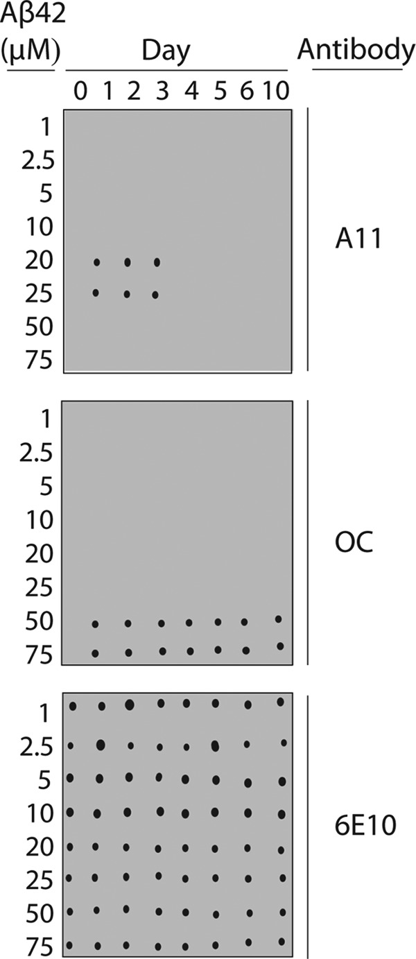
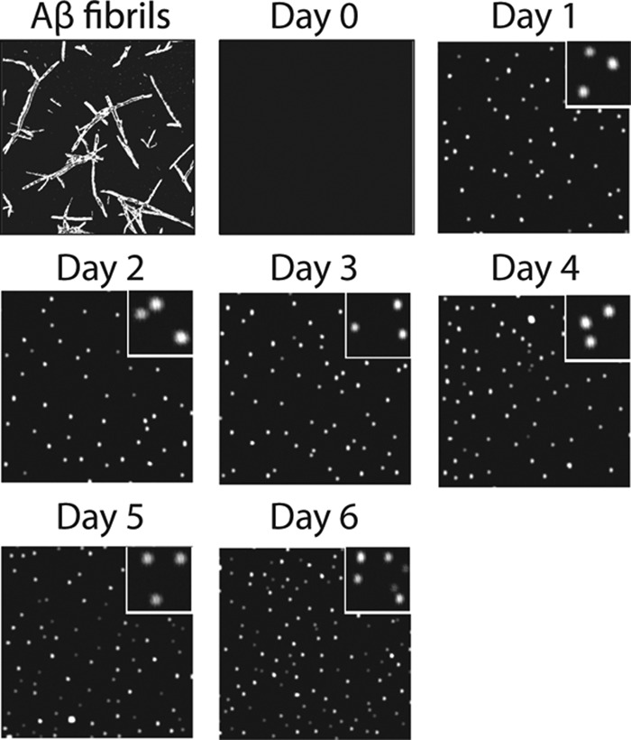
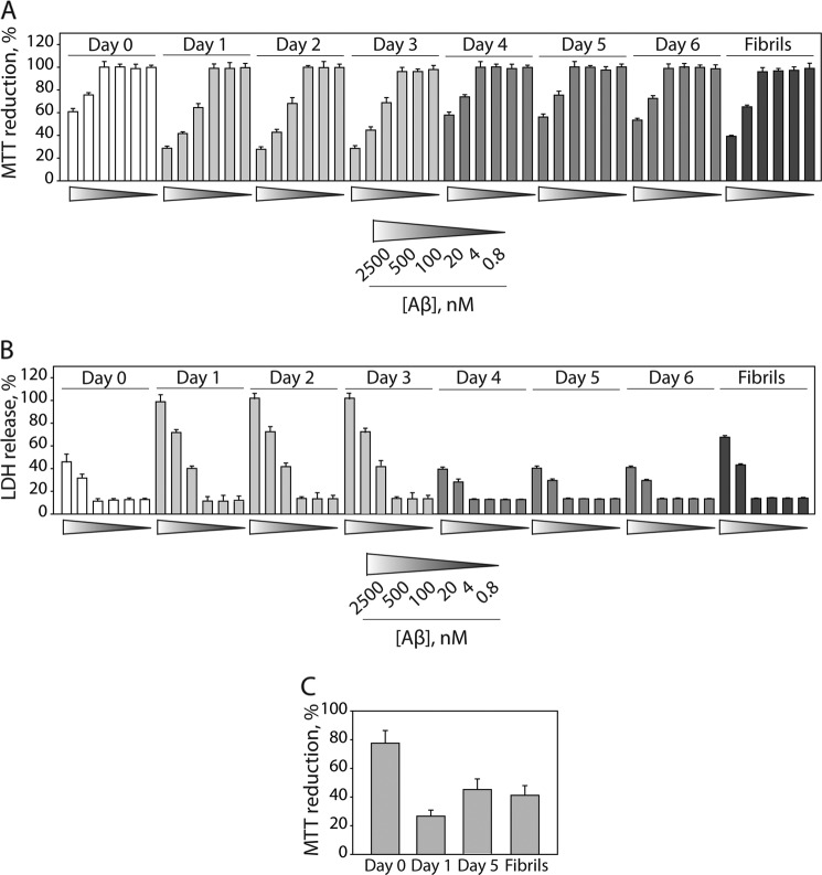
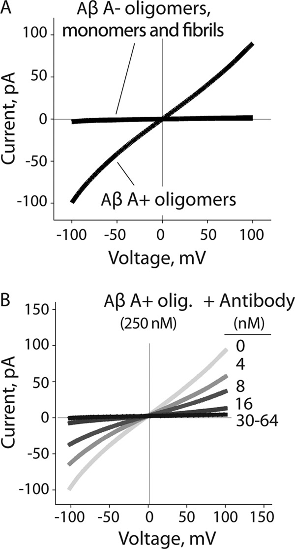
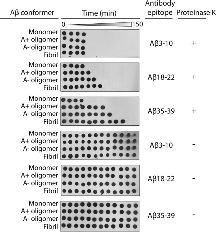
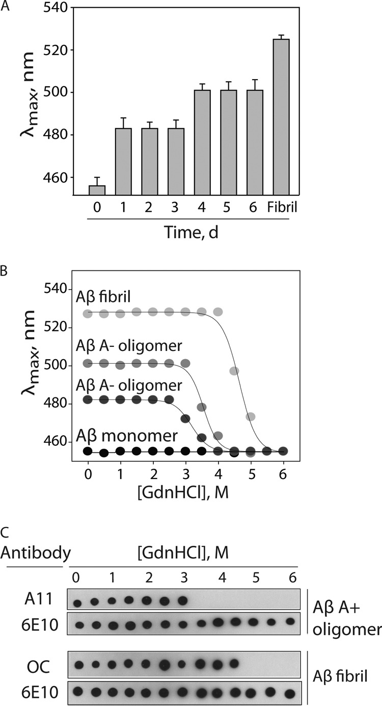
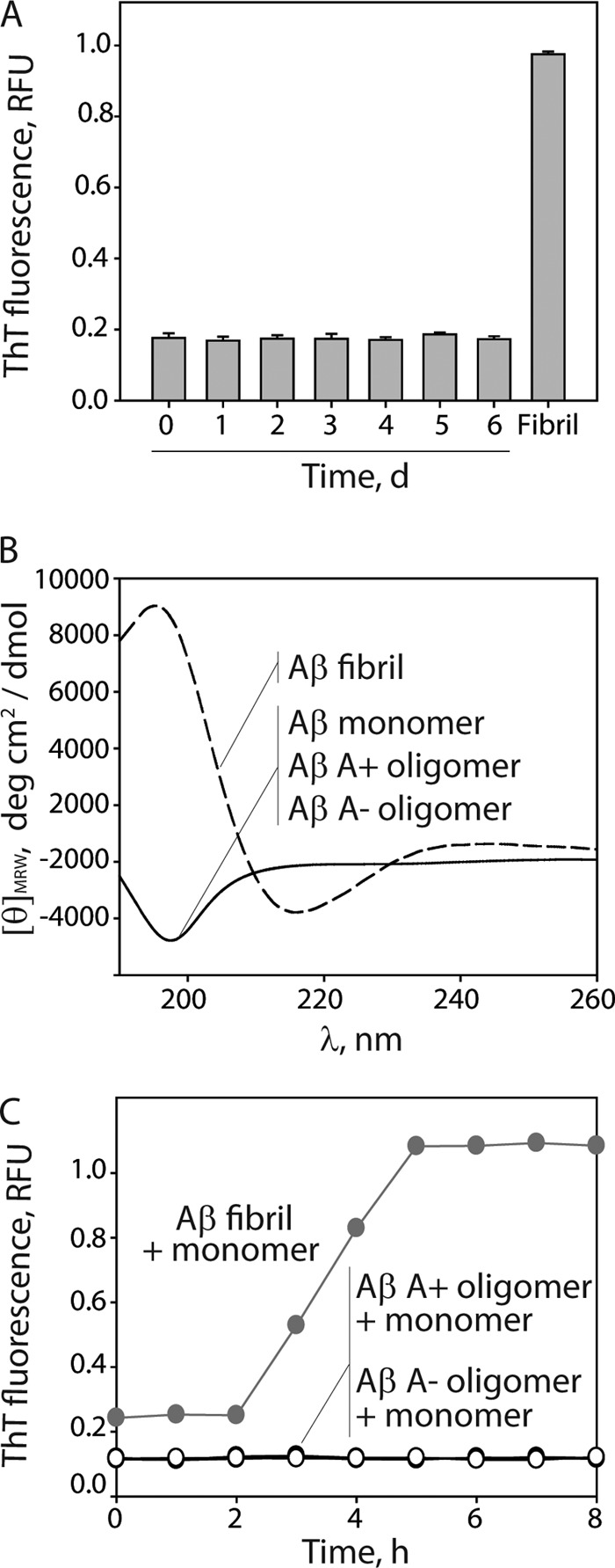
Several protein conformational disorders (Parkinson and prion diseases) are linked to aberrant folding of proteins into prefibrillar oligomers and amyloid fibrils. Although prefibrillar oligomers are more toxic than their fibrillar counterparts, it is difficult to decouple the origin of their dissimilar toxicity because oligomers and fibrils differ both in terms of structure and size. Here we report the characterization of two oligomers of the 42-residue amyloid β (Aβ42) peptide associated with Alzheimer disease that possess similar size and dissimilar toxicity. We find that Aβ42 spontaneously forms prefibrillar oligomers at Aβ concentrations below 30 μm in the absence of agitation, whereas higher Aβ concentrations lead to rapid formation of fibrils. Interestingly, Aβ prefibrillar oligomers do not convert into fibrils under quiescent assembly conditions but instead convert into a second type of oligomer with size and morphology similar to those of Aβ prefibrillar oligomers. Strikingly, this alternative Aβ oligomer is non-toxic to mammalian cells relative to Aβ monomer. We find that two hydrophobic peptide segments within Aβ (residues 16-22 and 30-42) are more solvent-exposed in the more toxic Aβ oligomer. The less toxic oligomer is devoid of β-sheet structure, insoluble, and non-immunoreactive with oligomer- and fibril-specific antibodies. Moreover, the less toxic oligomer is incapable of disrupting lipid bilayers, in contrast to its more toxic oligomeric counterpart. Our results suggest that the ability of non-fibrillar Aβ oligomers to interact with and disrupt cellular membranes is linked to the degree of solvent exposure of their central and C-terminal hydrophobic peptide segments.


Figures

FIGURE 1.
FIGURE 1.
Conformation-specific antibody analysis of Aβ assembly. Aβ42 conformers (1–75 μm) were assembled for 10 days (without agitation), and periodically deposited on nitrocellulose membranes. Afterward, the membranes were probed with conformation-specific (A11, prefibrillar oligomers (top), and OC, fibrillar conformers (middle)) and sequence-specific (6E10, N terminus of Aβ (bottom)) antibodies.
FIGURE 2.
FIGURE 2.
AFM analysis of Aβ oligomerization. Aβ42 (25 μm) was assembled without agitation, deposited on mica substrates, and imaged using AFM. Each image is 3 × 3 μm, and the inset images are 0.5 × 0.5 μm.
FIGURE 3.
FIGURE 3.
Toxicity analysis of Aβ oligomers and fibrils. Aβ42 (25 μm) was assembled without agitation and added to rat adrenal medulla cells (A and B) and rat primary cortical neuronal cells (C), and the relative toxicity was evaluated (n = 3). Error bars, S.D.
FIGURE 4.
FIGURE 4.
Impact of Aβ oligomers on lipid bilayer conductance. A, Aβ42 conformers (250 nm) were added to a reservoir on one side of the lipid bilayer (l-α-phosphocholine), and the membrane conductance was measured. B, Aβ A+ oligomers (12.5 μm) were mixed with the A11 antibody (0.2–3 μm) and diluted into a reservoir on one side of the lipid bilayer (50× dilution), and the membrane conductance was measured.
FIGURE 5.
FIGURE 5.
Rate of proteolytic fragmentation of Aβ peptide segments within Aβ oligomers. Aβ42 (25 μm) was incubated with Proteinase K (0.5 μg/ml), deposited periodically on nitrocellulose (every 10–30 min), and probed with antibodies specific for N-terminal (Aβ(3–10); 6E10), central (Aβ(18–22); 4G8), and C-terminal (Aβ(35–39); 9F1) Aβ epitopes. The time intervals were 0, 10, 20, 30, 40, 50, 60, 70, 80, 90, 120, and 150 min.
FIGURE 6.
FIGURE 6.
Analysis of hydrophobicity and conformational stability of Aβ oligomers. A, Aβ42 (25 μm) was assembled without agitation, and the hydrophobicity of Aβ conformers formed each day was evaluated using ANS fluorescence. The wavelength corresponding to the maximum ANS fluorescence is reported on the y axis. B and C, Aβ conformers (25 μm) were incubated with variable amounts of guanidine hydrochloride, and then their relative degree of unfolding was evaluated using ANS fluorescence (B) and antibodies (A11, prefibrillar oligomers; OC, fibrillar conformers; 6E10, N terminus of Aβ) (C). Error bars, S.D.
FIGURE 7.
FIGURE 7.
Characterization of the secondary structure and seeding activity of Aβ oligomers. Aβ42 (25 μm) was assembled without agitation (0–6 days), and its extent of fibrillization and secondary structure were evaluated using ThT fluorescence (A) and circular dichroism (B). C, Aβ fibrils and oligomers were mixed with Aβ monomers (25 μm, 5% seed), and their ThT fluorescence was monitored. RFU, relative fluorescence units. Error bars, S.D.

References

    1. Kirkitadze M. D., Bitan G., Teplow D. B. (2002) Paradigm shifts in Alzheimer's disease and other neurodegenerative disorders. The emerging role of oligomeric assemblies. J. Neurosci. Res. 69, 567–577 - PubMed
    1. Ono K., Condron M. M., Teplow D. B. (2009) Structure-neurotoxicity relationships of amyloid β-protein oligomers. Proc. Natl. Acad. Sci. U.S.A. 106, 14745–14750 - PMC - PubMed
    1. Hoshi M., Sato M., Matsumoto S., Noguchi A., Yasutake K., Yoshida N., Sato K. (2003) Spherical aggregates of β-amyloid (amylospheroid) show high neurotoxicity and activate Tau protein kinase I/glycogen synthase kinase-3β. Proc. Natl. Acad. Sci. U.S.A. 100, 6370–6375 - PMC - PubMed
    1. Cizas P., Budvytyte R., Morkuniene R., Moldovan R., Broccio M., Lösche M., Niaura G., Valincius G., Borutaite V. (2010) Size-dependent neurotoxicity of β-amyloid oligomers. Arch. Biochem. Biophys. 496, 84–92 - PMC - PubMed
    1. Sakono M., Zako T. (2010) Amyloid oligomers. Formation and toxicity of Aβ oligomers. FEBS J. 277, 1348–1358 - PubMed

Publication types