Review
. 2012 Nov;69(22):3715-37.
doi: 10.1007/s00018-012-0991-8. Epub 2012 May 1.

Current perspectives of the signaling pathways directing neural crest induction

Affiliations
Review

Current perspectives of the signaling pathways directing neural crest induction

Timothy J Stuhlmiller et al. Cell Mol Life Sci. 2012 Nov.

Abstract

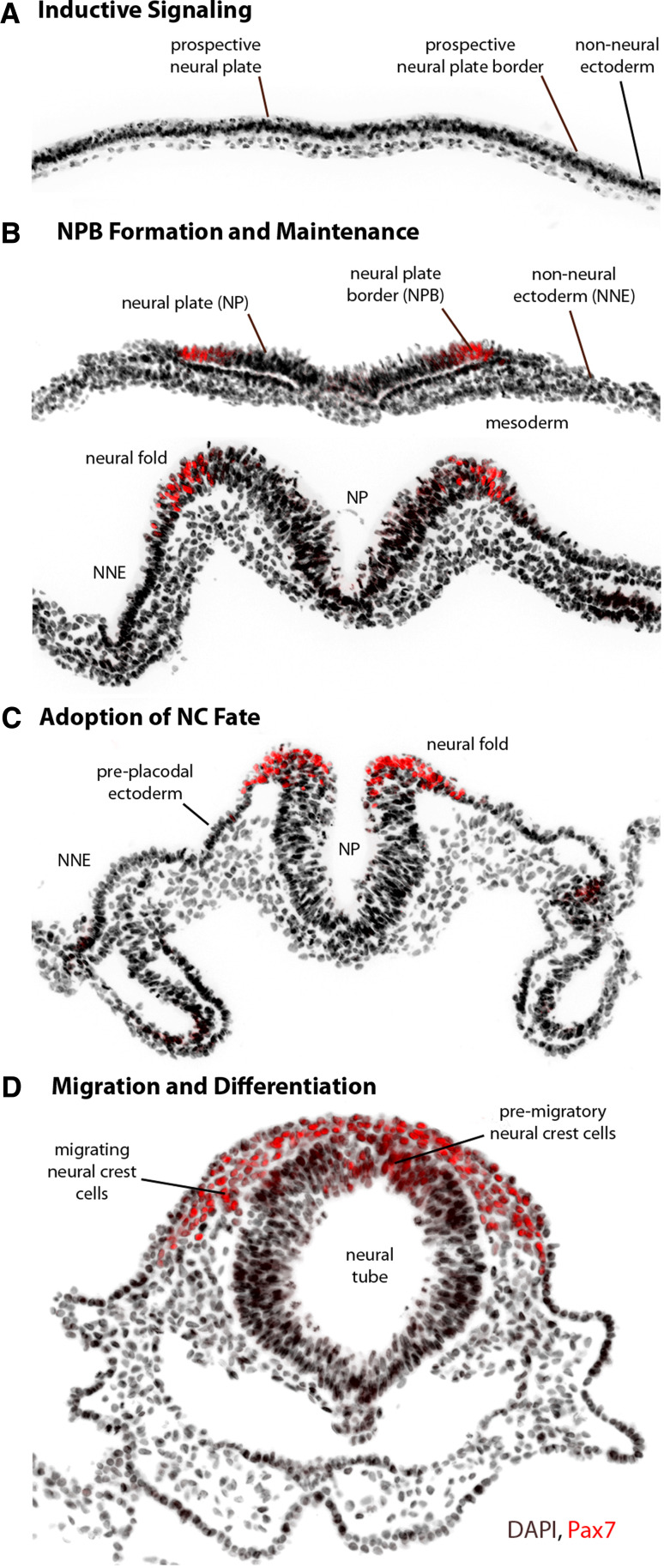
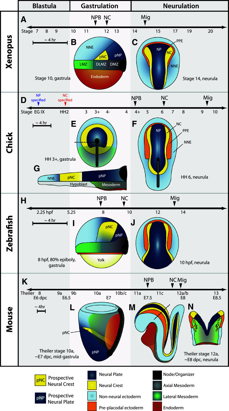
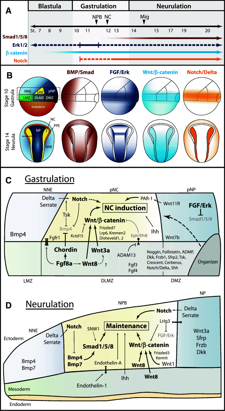
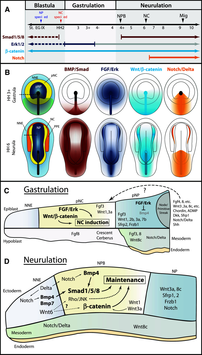
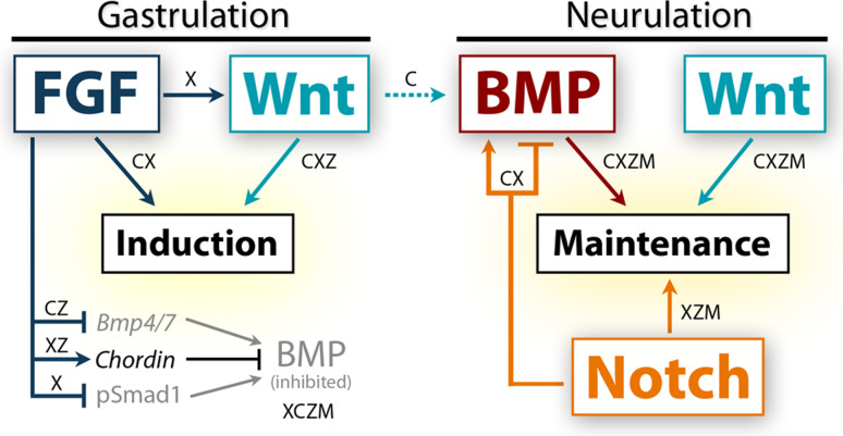
The neural crest is a migratory population of embryonic cells with a tremendous potential to differentiate and contribute to nearly every organ system in the adult body. Over the past two decades, an incredible amount of research has given us a reasonable understanding of how these cells are generated. Neural crest induction involves the combinatorial input of multiple signaling pathways and transcription factors, and is thought to occur in two phases from gastrulation to neurulation. In the first phase, FGF and Wnt signaling induce NC progenitors at the border of the neural plate, activating the expression of members of the Msx, Pax, and Zic families, among others. In the second phase, BMP, Wnt, and Notch signaling maintain these progenitors and bring about the expression of definitive NC markers including Snail2, FoxD3, and Sox9/10. In recent years, additional signaling molecules and modulators of these pathways have been uncovered, creating an increasingly complex regulatory network. In this work, we provide a comprehensive review of the major signaling pathways that participate in neural crest induction, with a focus on recent developments and current perspectives. We provide a simplified model of early neural crest development and stress similarities and differences between four major model organisms: Xenopus, chick, zebrafish, and mouse.


Figures

Fig. 1
Fig. 1
Morphogenesis and major events in early neural crest development. Images display major morphogenetic changes in the early stages of neural crest (NC) development from gastrulation to neurulation, using the chick embryo as an example. The neural plate border (NPB) and neural crest (NC) progenitors are marked by Pax7 in red. a Signaling molecules induce NC progenitors at the prospective NPB before and during the gastrula stage, but the source of inductive signals varies by organism. b NC progenitors are first identifiable with molecular markers of the neural plate border (NPB), including Msx1/2, Pax3/7, Zic1, Dlx3/5, Hairy2, Id3, and Ap2. The NPB is flanked medially by the neural plate (NP) and laterally by the non-neural ectoderm (NNE), with a layer of mesoderm found underneath. At the neurula stage, signaling between these tissues maintains the expression of NPB markers. c As the NP thickens and rises, the transcriptional activity of NPB specifiers and additional signaling events lead to the expression of NC specifiers at the neural folds, including Snail2, FoxD3, Sox9/10, Twist, cMyc, and Ap2. The pre-placodal ectoderm is found immediately lateral to the NC in rostral regions. d Soon after the NC fate is established, NC cells undergo an epithelial-to-mesenchymal transition and migrate throughout the body and differentiate into a multitude of derivatives. In the chick, NC cells migrate soon after the neural tube fuses, but in most other organisms, NC cells begin to migrate before the neural tube is closed
Fig. 2
Fig. 2
Timing and morphology of early neural crest development in Xenopus, chick, zebrafish, and mouse. a, d, h, k Timelines for early events in NC development. Note the appearance of neural plate border markers (NPB) and neural crest specifiers (NC) occurs during gastrulation in anamniotes (Xenopus and zebrafish) and after gastrulation in amniotes (chick and mouse). Anamniotes progress at a higher rate of development and the time between events is generally very short—compare sizes of ~4-h time bars. a In Xenopus, markers of the neural plate border are first apparent at Nieuwkoop and Faber stage 11.5 and immediately precede expression of neural crest specifiers at stage 12, before the end of gastrulation. NC migration (Mig) begins around stage 15. b Lateral view of early Xenopus gastrula. Animal pole is up, dorsal to the right. Prospective neural crest tissue (pNC) is found above the dorsolateral marginal zone (DLMZ), based on fate-mapping studies [31]. LMZ lateral marginal zone, DMZ dorsal marginal zone. c Dorsal view of a Xenopus neurula. Anterior is up. d In the chick, neural tissue is specified before the egg is laid at Eyal-Giladi (EG) stage IX, while neural crest tissue is specified by Hamburger and Hamilton (HH) stage 2. Markers of the neural plate border are not apparent until after gastrulation at stage 4+. The first neural crest specifiers are not expressed until stage 6. Migration begins between stage 9 and 10. e Dorsal view of mid-gastrula. Prospective neural crest tissue is found in a ring around the prospective neural plate (pNP) until post-gastrula stages when the anterior NPB is specified to become pre-placodal ectoderm [30]. f Lateral section through the dotted line in e. At pre-gastrula and early gastrula stages, the prospective neural crest is situated above the hypoblast, an extra-embryonic tissue. As mesoderm and endoderm ingress, the hypoblast is displaced anteriorly, and by the end of gastrulation prospective neural crest tissue is underlain by mesoderm. g Dorsal view of neurula, anterior is up. NC specifiers are initially only expressed in the anterior-most aspect of the neural folds. h In the zebrafish, neural plate border markers and neural crest specifiers are first expressed during gastrulation. Migration occurs after 13 h post-fertilization (hpf). i Lateral view of zebrafish gastrula. Animal pole is up, dorsal to the right. Location of prospective neural crest is inferred from expression of Msxb [70] and Pax3 [113]. j Dorsal view of neurula, anterior is up. k In the mouse, most neural plate border markers are first detectable around E7.5. Neural crest specifiers are expressed by E7.75, and NC cells begin migrating almost immediately after this expression. Listed below the timeline are approximate stages by Theiler stage, and embryonic days post coitum (dpc). l Lateral view of mouse gastrula. Anterior to the left. The mouse embryo develops with the prospective ectoderm as the interior layer. Location of the prospective neural crest is inferred from the position of prospective neural and non-neural tissues, and the expression of NPB markers by E7.5. m Lateral view of neurula. Anterior to the left. n Section through the dotted line in m. Although the neural tube has not yet closed, NC cells are migrating extensively
Fig. 3
Fig. 3
Temporal and spatial participation of signaling molecules involved in Xenopus neural crest induction. a Timeline of signaling pathway activation and requirement in early NC development. Closed arrows/lines indicate activation and requirement in NC tissues. Dotted lines indicate activation in NC tissues, but requirement is unknown. BMP/Smad signaling must be inhibited during gastrulation, but activated upon neurulation. The specific stage when Smad signaling first becomes activated in the NC has not been determined. FGF/Erk signaling is activated throughout early NC development, but has only been functionally implicated during early gastrula stages [49, 50]. Wnt/β-catenin signaling is thought to be required at all stages of early NC development, but becomes more strongly activated by neurulation [31]. The precise time when Notch is required is still debated, but may play a role in the initial induction during gastrulation. b Spatial activation of BMP, FGF, Wnt, and Notch signaling during Xenopus gastrulation and neurulation. BMP/Smad, FGF/Erk, and Wnt/β-catenin activation based on data from [199, 200]. Notch/Delta activation inferred from requirements in germ layer segregation and NC development [8, 12, 201]. Overall, spatiotemporal activation of these pathways is conserved between Xenopus and zebrafish. c Spatial expression and participation of signaling molecules in Xenopus neural crest induction at the gastrula stage. Diagram corresponds to dotted box of stage 10 gastrula in b. Molecules in bold have support from multiple studies. Solid lines indicate known relationships, dotted lines indicate potential relationships. NC induction results from the combined action of Wnt/β-catenin, FGF, Indian Hedgehog, and non-canonical Wnt signaling. Fgf8a is thought to regulate the expression of Wnt8 in the dorsolateral marginal zone (DLMZ), but may signal to the prospective neural crest itself. Wnt8 and Wnt3a signaling from the DLMZ activate canonical Wnt signaling in the prospective neural crest. Multiple agonists and antagonists of BMP and Wnt signaling are expressed in the Organizer, DMZ, and DLMZ and function in dorsal–ventral and anterior–posterior patterning, and these molecules likely also participate in NC induction (dotted line). Expression of other potential signaling molecules and regulators is presented. See main text for details on the participation of individual signaling molecules. d Participation of signaling molecules in the maintenance of NC progenitors in Xenopus neurulation. Diagram corresponds to section at dotted line in stage 14 neurula in b. NC maintenance requires activation of Smad1/5/8, Wnt/β-catenin, Notch/Delta, Indian Hedgehog, and Endothelin-A signaling. BMP and Wnt signals are likely mediated by Bmp4, Bmp7, Wnt1, and Wnt8, expressed in the neural folds upon neurulation. Additionally, Wnt8 is present in the paraxial mesoderm and Wnt3a in the neural plate. Notch signaling is thought to operate both by regulating Bmp ligand levels and leading to the expression of NC specifiers. See main text for details on the participation of individual signaling molecules. Expression data gathered from references in text and from http://www.xenbase.org
Fig. 4
Fig. 4
Temporal and spatial participation of signaling pathways involved in chick neural crest induction. a Timeline of signaling pathway activation and requirement in early NC development. Closed arrows/lines indicate activation and requirement in NC tissues. Dotted lines indicate activation in NC tissues, but requirement is unknown. Smad1/5/8 signaling is active in the entire epiblast at blastula stages, but is inactivated by gastrulation [75]. Signaling becomes active with the expression of NPB markers, and remains active through to migration. Erk signaling is also active in most of the epiblast at blastula stages, and is required for neural induction until gastrulation. A requirement for FGF/Erk signaling in NC induction was only demonstrated during gastrulation. Erk signaling remains active in the NPB and NC tissues through to migration, but is no longer required for NC development (gray line). Wnt/β-catenin signaling is thought to be necessary for all stages of early NC development. A requirement for Notch/Delta signaling was demonstrated at mid-neurula stages. b Spatial activation of BMP, FGF, Wnt, and Notch signaling during chick gastrulation and neurulation. BMP/Smad activation based on [76]. FGF/Erk activation based on [76, 128]. Wnt/β-catenin activation inferred from expression of agonists and antagonists, and functional requirements for Wnt signaling. Notch/Delta activation is based on expression of molecules and functional requirements [154]. c Spatial expression of relevant signaling molecules and requirements for chick neural crest induction during gastrulation. Diagram corresponds to section at dotted line HH 3+ gastrula in b. Functional studies have demonstrated a requirement for FGF/Erk and Wnt/β-catenin signaling, but the participation of specific signaling molecules has not been challenged. The spatial expression of some potential signaling molecules is presented. Multiple FGF and Wnt agonists and BMP and Wnt antagonists are expressed in the node/primitive streak, but it is unclear whether these molecules can diffuse the distance to influence the prospective NC tissue (dotted arrow). d Spatial expression and participation of signaling molecules and pathways in the maintenance of NC progenitors during chick neurulation. Diagram corresponds to section at dotted line in HH 6 neurula in b. Smad1/5/8 and β-catenin are likely activated by Bmp4, Bmp7, Wnt1, and Wnt3a expressed in the neural folds and adjacent NNE. Wnt6 in the NNE has also been implicated in NC development, but may act through the non-canonical Rho/JNK pathway. Notch signaling likely participates indirectly by regulating Bmp4 expression. Spatial expression of other potential signaling molecules is presented. Expression data gathered from references in the text and from http://geisha.arizona.edu/geisha
Fig. 5
Fig. 5
A model of signaling participation during the two phases of neural crest induction. In this figure, we present a simplified model of the major signaling events thought to occur in NC induction, drawing on evidence from all four of the organisms discussed. Since the precise time, source, and cross-regulation between pathways vary between species, the model organism is noted where a specific interaction or activity is known to occur. X Xenopus, C chick, Z zebrafish, M mouse. During gastrulation, FGF and Wnt signaling are both known to induce the neural crest at the prospective NPB, activating the expression of NPB specifiers. Xenopus studies demonstrate that FGF regulates Wnt signaling during this first phase, but evidence from chick and Xenopus suggests FGF acts directly as well. BMP signaling must be at least partially inhibited for this first step, and FGF participates in BMP attenuation on multiple levels. The transition to the second phase involves the activation of BMP signaling, and research on chick explants suggests Wnts may participate in this BMP activation. Since FGF contributes to BMP antagonism before and during gastrulation, the restriction of FGF activity or insensitivity of the NPB to FGF signals also likely plays a role in this transition. In the second phase, BMP and Wnt signaling converge to maintain the expression of NPB specifiers and initiate the expression of NC specifiers. Notch signaling is known to refine the domain of BMP activity, but some evidence suggests Notch acts directly on the neural crest population as well. Throughout later neural crest development, these signaling pathways continue to participate in migration and differentiation

References

    1. Meulemans D, Bronner-Fraser M. Gene-regulatory interactions in neural crest evolution and development. Dev Cell. 2004;7(3):291–299. - PubMed
    1. Sauka-Spengler T, Bronner-Fraser M. A gene regulatory network orchestrates neural crest formation. Nat Rev Mol Cell Biol. 2008;9(7):557–568. - PubMed
    1. Hong CS, Saint-Jeannet JP. The activity of Pax3 and Zic1 regulates three distinct cell fates at the neural plate border. Mol Biol Cell. 2007;18(6):2192–2202. - PMC - PubMed
    1. Monsoro-Burq AH, Wang E, Harland R. Msx1 and Pax3 cooperate to mediate FGF8 and WNT signals during Xenopus neural crest induction. Dev Cell. 2005;8(2):167–178. - PubMed
    1. Sato T, Sasai N, Sasai Y. Neural crest determination by co-activation of Pax3 and Zic1 genes in Xenopus ectoderm. Development. 2005;132(10):2355–2363. - PubMed