. 2012 May 7;209(5):935-45.
doi: 10.1084/jem.20112583. Epub 2012 Apr 30.

Resident CD141 (BDCA3)+ dendritic cells in human skin produce IL-10 and induce regulatory T cells that suppress skin inflammation

Affiliations

Resident CD141 (BDCA3)+ dendritic cells in human skin produce IL-10 and induce regulatory T cells that suppress skin inflammation

Chung-Ching Chu et al. J Exp Med. .

Abstract

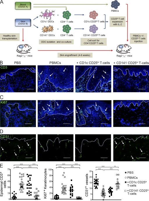
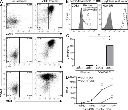
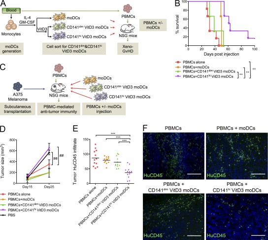
Human skin immune homeostasis, and its regulation by specialized subsets of tissue-residing immune sentinels, is poorly understood. In this study, we identify an immunoregulatory tissue-resident dendritic cell (DC) in the dermis of human skin that is characterized by surface expression of CD141, CD14, and constitutive IL-10 secretion (CD141(+) DDCs). CD141(+) DDCs possess lymph node migratory capacity, induce T cell hyporesponsiveness, cross-present self-antigens to autoreactive T cells, and induce potent regulatory T cells that inhibit skin inflammation. Vitamin D(3) (VitD3) promotes certain phenotypic and functional properties of tissue-resident CD141(+) DDCs from human blood DCs. These CD141(+) DDC-like cells can be generated in vitro and, once transferred in vivo, have the capacity to inhibit xeno-graft versus host disease and tumor alloimmunity. These findings suggest that CD141(+) DDCs play an essential role in the maintenance of skin homeostasis and in the regulation of both systemic and tumor alloimmunity. Finally, VitD3-induced CD141(+) DDC-like cells have potential clinical use for their capacity to induce immune tolerance.


Figures

Figure 1.
Figure 1.
Identification and characterization of skin migratory CD141+ DDCs. (A and B) Healthy human skin DDCs were stained for CD1c, CD11c, CD14, and CD1a. (C) CD141+HLA-DR+ DDCs staining in the upper dermis of normal human skin (arrows). Dashed line indicates epidermal–dermal junction. (D) Filamentous actin (F-actin) staining of cell-sorted CD141+ DDCs. (E) CD141+ DDCs or CD1c+ DDCs stained for CD83, CD80, CD86, HLA-ABC, HLA-DR, and MMR. (F) Human CD45+ staining of lymph nodes and spleens of BALB/c Rag2−/−γc−/− mice bearing human skin transplants (Tx). (G) Human CD141+ and CD3 staining in lymph nodes of Tx mice. (H) mRNA expression for human CD141, cyclophilin, and mouse GAPDH in spleens and lymph nodes of Tx mice. Blood and skin CD141+ DCs expression of BATF3, Necl2, CLEC9A, and XCR1 (I) and CD141+ DDCs expression of CLEC9A (J) as compared with CD1c+ DDCs. (K) CD141+ DDCs or CD1c+ DDCs pulsed with PPI were incubated with autoreactive CD8+ T cell clones and analyzed for proliferation. Net CPM represents proliferation of the clone in pulsed DDC co-culture minus nonpulsed DDC co-culture. Error bars indicate SEM. Results are representative of at least 30 (A), at least three (B–E), and two (F–K) independent experiments. Two-way ANOVA test; *, P < 0.05.
Figure 2.
Figure 2.
CD141+ DDCs are immunoregulatory. (A) CD141+ DDCs or CD1c+ DDCs expression of ILT3 and (B) production of IL-10 in the absence or presence of CD40 ligand-transfected L cells (CD40L Tx). (C) CD141+ DDCs or CD1c+ DDCs stimulation of allogeneic CD4+ T cell proliferation. (D) CD141+ DDCs or CD1c+ DDCs co-cultured with allogeneic CD4+ T cells in the presence and absence of anti–IL-10 receptor antibody. CD4+CD25hi T cells induced by CD1c+ DDCs (CD1c CD25hi T cells) or CD141+ DDCs (CD141 CD25hi T cells) stained for surface CTLA-4 (E) and proliferation in response to alloantigen stimulation (F). (G) IL-2–expanded CD25hi T cells proliferative response to secondary polyclonal stimulation and (H) suppression of CD4+CD25 T eff cell proliferation. Error bars indicate SEM. Results are representative of five (A–C, E, and F) and two (D, G, and H) independent experiments. Wilcoxon matched pairs test (A), one-way ANOVA (B), two-way ANOVA (C, F, and H), or unpaired Student’s t test (D, G). *, P < 0.05; **, P < 0.01; ***, P < 0.001; #, P < 0.05.
Figure 3.
Figure 3.
CD141+ DDC-induced CD25hi T reg cells inhibit human alloimmune cell–mediated skin inflammation. (A) Experimental strategy to assess CD25hi T cell suppression. BALB/c Rag2−/−γc−/− mice transplanted with human skin were injected with PBS, allogeneic PBMCs alone, or in combination with CD141 CD25hi T cells or CD1c CD25hi T cells. Representative fields from stained skin grafts for epidermal CD3+ T cell infiltration (B, arrows), epidermal keratinocyte expression of the proliferation marker Ki67 (C, arrows), and intact human CD31 superficial dermal microvasculature (D, arrows). Dashed lines indicate epidermal–dermal junction. Nuclei are stained with DAPI (blue). Bars, 100 µm. (E) Quantitative histological analysis of at least three independent visual fields per skin graft. Lines represent the mean (n = 5–9 animals per treatment group). Results are combined data from three independent experiments. One-way ANOVA test; **, P < 0.01; ***, P < 0.001.
Figure 4.
Figure 4.
VitD3 induces CD141+ DDC-like phenotype in blood DCs. (A) VitD3-treated blood CD1c+ DC expression of CD141 with CD14, ILT3, and MMR. VitD3-induced CD141+ DCs or VitD3-induced CD141 DCs were assessed for their expression of CD83 and HLA-DR in the presence of a cytokine maturation cocktail (B), production of IL-10 in the presence and absence of CD40L Tx stimulation (C), and ability to stimulate allogeneic CD4+ T cell proliferation (D). Results are representative of at least 12 (A) and 3 (B–D) independent experiments. One-way ANOVA (C) or two-way ANOVA (D); **, P < 0.01; ##, P < 0.01.
Figure 5.
Figure 5.
VitD3-induced CD141hi DCs suppress xeno-GvHD and antitumor immunity. (A) Strategy for preclinical assessment of adoptive cell therapy in xeno-GvHD. (B) NSG mice were injected with either PBMCs alone or in combination with moDCs, CD141dim VitD3 moDCs, or CD141hi VitD3 moDCs to induce xeno-GVHD. Survival was monitored over time. (C) Strategy to determine regulation of antitumor alloimmunity. Tumor size (D) and CD45+ infiltration (E and F) were assessed in animals injected with PBMCs alone or in combination with moDCs, CD141dim VitD3 moDCs or CD141hi VitD3 moDCs to induce antitumor alloimmunity (E) Quantitative histological analysis of tumor CD45+ infiltration from at least three independent visual fields per skin graft. Lines represent the mean (n = 3–6 animals per treatment group). (F) Representative pictures of human CD45+ infiltrates in tumors. Nuclei are stained with DAPI (blue). Bars, 100 µm. Results are pooled from three independent experiments (n = 3–6 animals per treatment group). One-way ANOVA (E), two-way ANOVA (D), or Mantel-Cox (B) test; **, P < 0.01; ***, P < 0.001; ##, P < 0.01.

References

    1. Adorini L., Giarratana N., Penna G. 2004. Pharmacological induction of tolerogenic dendritic cells and regulatory T cells. Semin. Immunol. 16:127–134 10.1016/j.smim.2003.12.008 - DOI - PubMed
    1. Anas A., van der Poll T., de Vos A.F. 2010. Role of CD14 in lung inflammation and infection. Crit. Care. 14:209 10.1186/cc8850 - DOI - PMC - PubMed
    1. Azukizawa H., Döhler A., Kanazawa N., Nayak A., Lipp M., Malissen B., Autenrieth I., Katayama I., Riemann M., Weih F., et al. 2011. Steady state migratory RelB+ langerin+ dermal dendritic cells mediate peripheral induction of antigen-specific CD4+ CD25+ Foxp3+ regulatory T cells. Eur. J. Immunol. 41:1420–1434 10.1002/eji.201040930 - DOI - PubMed
    1. Bachem A., Güttler S., Hartung E., Ebstein F., Schaefer M., Tannert A., Salama A., Movassaghi K., Opitz C., Mages H.W., et al. 2010. Superior antigen cross-presentation and XCR1 expression define human CD11c+CD141+ cells as homologues of mouse CD8+ dendritic cells. J. Exp. Med. 207:1273–1281 10.1084/jem.20100348 - DOI - PMC - PubMed
    1. Bedoui S., Whitney P.G., Waithman J., Eidsmo L., Wakim L., Caminschi I., Allan R.S., Wojtasiak M., Shortman K., Carbone F.R., et al. 2009. Cross-presentation of viral and self antigens by skin-derived CD103+ dendritic cells. Nat. Immunol. 10:488–495 10.1038/ni.1724 - DOI - PubMed

Publication types

MeSH terms