. 2012 May 15;109(20):7888-92.
doi: 10.1073/pnas.1205869109. Epub 2012 Apr 30.

Calpain cleaves and activates the TRPC5 channel to participate in semaphorin 3A-induced neuronal growth cone collapse

Affiliations

Calpain cleaves and activates the TRPC5 channel to participate in semaphorin 3A-induced neuronal growth cone collapse

J Stefan Kaczmarek et al. Proc Natl Acad Sci U S A. .

Abstract

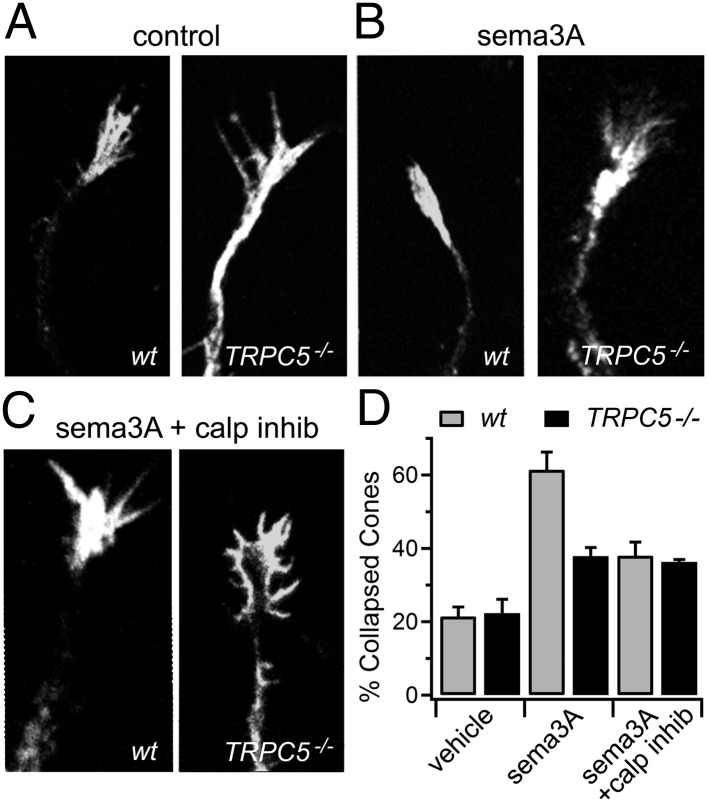
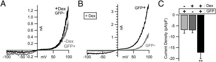
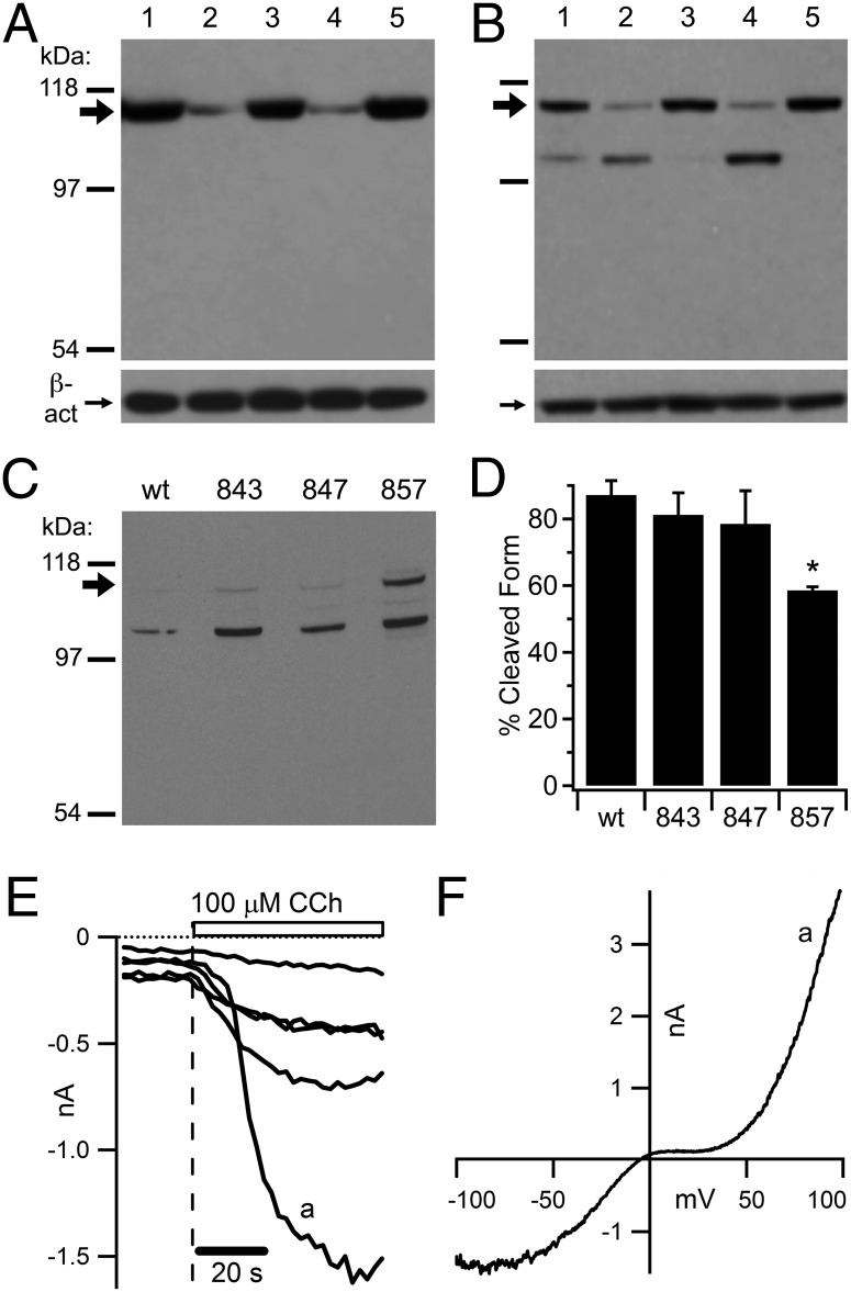
The nonselective cation channel transient receptor potential canonical (TRPC)5 is found predominantly in the brain and has been proposed to regulate neuronal processes and growth cones. Here, we demonstrate that semaphorin 3A-mediated growth cone collapse is reduced in hippocampal neurons from TRPC5 null mice. This reduction is reproduced by inhibition of the calcium-sensitive protease calpain in wild-type neurons but not in TRPC5(-/-) neurons. We show that calpain-1 and calpain-2 cleave and functionally activate TRPC5. Mutation of a critical threonine at position 857 inhibits calpain-2 cleavage of the channel. Finally, we show that the truncated TRPC5 predicted to result from calpain cleavage is functionally active. These results indicate that semaphorin 3A initiates growth cone collapse via activation of calpain that in turn potentiates TRPC5 activity. Thus, TRPC5 acts downstream of semaphorin signaling to cause changes in neuronal growth cone morphology and nervous system development.


Conflict of interest statement

The authors declare no conflict of interest.

Figures

Fig. 1.
Fig. 1.
TRPC5 knockout and calpain inhibition reduce sema3A-induced hippocampal growth cone collapse. (A) Untreated, cultured hippocampal neuronal growth cones from WT and TRPC5 null (Trpc5−/−) mice, fixed and stained with the actin-binding peptide phalloidin conjugated to Alexa Fluor 568. (B) Growth cones from neurons treated for 5 min with 1 nM sema3A before fixation. (C) Growth cones from neurons preincubated with the calpain inhibitors calpain inhibitor III (5 μM) and calpeptin (10 μM) for 30 min and then treated with sema3A. (D) Growth cones from the indicated genotypes and treatments were counted and the collapsed fraction quantified (three coverslips from two separate experiments; n = 98–100 cones per condition). Ambiguous growth cones not closely resembling the examples presented were excluded from analysis. P values in text are calculated using Student t test.
Fig. 2.
Fig. 2.
Coexpression of constitutively active calpain-2 S50E with TRPC5 increases basal channel currents. (A and B) Representative whole-cell current–voltage (I-V) relationships from HEK cells stably expressing TRPC5 cotransfected with calpain-2 S50E under the control of a dexamethasone-inducible promoter, CAPNS1, and eGFP as a marker. −Dex, uninduced; +Dex, dexamethasone-treated cells; GFP−, GFP-negative cells; GFP+, GFP-positive cells. The pipette contained 5 mM HEDTA and 5 μM free Ca2+ to accentuate basal TRPC5 currents. The bath contained the standard 2 mM Ca2+ extracellular solution. I-V relationships were obtained shortly after break-in (<1 min). Both traces in B are from dexamethasone-treated cells. (C) Quantification of whole-cell current density at −100 mV from uninduced, GFP-positive (light gray column; n = 10), induced but GFP-negative (dark gray column; n = 10), and induced, GFP-positive (black column, n = 10) cells. **P < 0.01 (Student t test).
Fig. 3.
Fig. 3.
Purified calpains activate TRPC5 channels in excised patches. Average single channel conductance observed for TRPC5 was 39.5 ± 0.5 pS (n = 20). (A, Upper) Activity (NPO) of TRPC5 channels in a representative inside-out excised patch from a HEK cell stably expressing mTRPC5, activated by 15 μg/mL purified calpain-1 (indicated by dashed line and open bar), collected in 30-s bins. (A, Lower) Currents at the time points indicated by the small letters above showing individual TRPC5 channel openings at the indicated voltage. C, closed (no open channels); O or 1, one open channel; 2, two open channels. We used an intracellular solution with 5 mM HEDTA and 5 μM free Ca2+ throughout the experiment. (B) Same as A, except that we perfused purified calpain-1 plus a protease inhibitor mixture including the cysteine protease inhibitor E-64 (14 μM). (C) Same as A, except that we perfused purified calpain-2 onto the patch and used an intracellular solution containing 2 mM Ca2+. (D) Same as A, except that we perfused calpain-2, which had been phosphorylated in vitro with constitutively active MAPK1, onto the patch in an intracellular solution containing 100 nM free Ca2+. (E) Same as D, except that MAPK1 was boiled before incubation with calpain-2 before the experiment. (F) Quantification of the effects of purified calpains on TRPC5. Fold increase is the ratio between the NPO of the peak bin following treatment, divided by the average NPO preceding treatment. Each patch is indicated by its own marker; the solid bar is the mean. *P < 0.05; **P < 0.01 ( Student t test; n = 6–8 for each experiment). Burst activity shown as rising and falling NPO is characteristic of TRPC5.
Fig. 4.
Fig. 4.
Calpain-1 and calpain-2 cleave TRPC5 at its C terminus, generating a fragment likely to retain ion channel function, and this cleavage is significantly reduced by mutation of threonine 857 to alanine. (A and B) We briefly sonicated HEK cells stably expressing an N-terminally HA-tagged mTRPC5 protein and treated the resulting homogenates with 5 μg/mL purified calpain-1 (lane 2), calpain-1 plus 10 μM calpeptin and 5 μM calpain inhibitor III (lane 3), 5 μg/mL purified calpain-2 (lane 4), or calpain-2 plus the aforementioned inhibitors (lane 5) in a 2 mM Ca2+ buffer (control untreated; lane 1). (A, Upper) Western blot of calpain-treated HA-TRPC5 probed with a TRPC5 C-terminal monoclonal antibody. The large arrow below the 118-kDa marker indicates full-length HA-TRPC5 protein. (B, Upper) Western blot of calpain-treated HA-TRPC5 probed with an HA monoclonal antibody. (A, Lower; and B, Lower) A monoclonal antibody to β-actin shows that equal protein was loaded. (C) Western blot of calpain-2 treated homogenates from HEK cells transfected with an HA-TRPC5 wild-type construct or constructs containing the point mutations F843A, G847A, and T857A probed with anti-HA. The large arrow below the 118-kDa marker indicates full-length HA-TRPC5 protein. (D) Quantification of TRPC5 present in the cleaved form from the blot in C. *P < 0.05 (Student t test; n = 2 blots from independent experiments). (E and F) A modified plasmid containing mTRPC5 truncated at N854 was coexpressed with the muscarinic-type 1 receptor (M1R) and eGFP in HEK cells. (E) Representative whole-cell currents recorded at −100 mV; application of the M1R agonist carbachol at 100 μM (vertical dashed line and open bar) significantly increased current density in five GFP-positive cells. The pipette contained a solution with 5 mM HEDTA and 5 μM free Ca2+ to potentiate any TRPC5-like currents. (F) Current–voltage relationship (I-V) taken at the point indicated by a in E.

References

    1. O’Donnell M, Chance RK, Bashaw GJ. Axon growth and guidance: Receptor regulation and signal transduction. Annu Rev Neurosci. 2009;32:383–412. - PMC - PubMed
    1. Hong K, Nishiyama M, Henley J, Tessier-Lavigne M, Poo M. Calcium signalling in the guidance of nerve growth by netrin-1. Nature. 2000;403:93–98. - PubMed
    1. Sakai T, et al. Mouse semaphorin H induces PC12 cell neurite outgrowth activating Ras-mitogen-activated protein kinase signaling pathway via Ca2+ influx. J Biol Chem. 1999;274:29666–29671. - PubMed
    1. To KC, Church J, O’Connor TP. Combined activation of calpain and calcineurin during ligand-induced growth cone collapse. Mol Cell Neurosci. 2007;36:425–434. - PubMed
    1. Gerhold KA, Bautista DM. Molecular and cellular mechanisms of trigeminal chemosensation. Ann N Y Acad Sci. 2009;1170:184–189. - PMC - PubMed

Publication types