. 2012 Jun 5;109(23):9101-6.
doi: 10.1073/pnas.1206069109. Epub 2012 Apr 30.

One influenza virus particle packages eight unique viral RNAs as shown by FISH analysis

Affiliations

One influenza virus particle packages eight unique viral RNAs as shown by FISH analysis

Yi-ying Chou et al. Proc Natl Acad Sci U S A. .

Abstract

Influenza A virus possesses a segmented genome of eight negative-sense, single-stranded RNAs. The eight segments have been shown to be represented in approximately equal molar ratios in a virus population; however, the exact copy number of each viral RNA segment per individual virus particles has not been determined. We have established an experimental approach based on multicolor single-molecule fluorescent in situ hybridization (FISH) to study the composition of viral RNAs at single-virus particle resolution. Colocalization analysis showed that a high percentage of virus particles package all eight different segments of viral RNAs. To determine the copy number of each RNA segment within individual virus particles, we measured the photobleaching steps of individual virus particles hybridized with fluorescent probes targeting a specific viral RNA. By comparing the photobleaching profiles of probes against the HA RNA segment for the wild-type influenza A/Puerto Rico/8/34 (PR8) and a recombinant PR8 virus carrying two copies of the HA segment, we concluded that only one copy of HA segment is packaged into a wild type virus particle. Our results showed similar photobleaching behaviors for other RNA segments, suggesting that for the majority of the virus particles, only one copy of each RNA segment is packaged into one virus particle. Together, our results support that the packaging of influenza viral genome is a selective process.


Conflict of interest statement

The authors declare no conflict of interest.

Figures
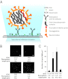
Fig. 1.
Fig. 1.
(A) Schematic for FISH analysis on single-virus particle. Viral RNA hybridized with multiple fluorophore-labeled probes is visualized using TIRF microscopy. Biotinylated antibodies against the influenza virus surface protein HA are immobilized on imaging surface passivated with PEG and supplemented with biotinylated PEG and NeutrAvidin. Influenza virus particles are captured onto the imaging surface with the immobilized anti-HA antibodies. FISH is performed on virus particles that are captured, fixed, and permeabilized on the imaging surface. (B) TIRF images of FISH signals from hybridization reactions using a mixture of 48 DNA probes singly labeled with fluorophores targeting viral RNA on a blank slide (no immobilized virus), PR8 virus-immobilized slides, and FISH signals from hybridization reactions on PR8 virus-immobilized slide using 48 Cy3-labled probes against the RIG-I mRNA are shown. (C) Average number of fluorescent spots per 1,000 μm2 is shown (number of fields, n > 10).
Fig. 2.
Fig. 2.
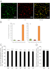
Copackaging of different viral RNA segments into a virus particle. (A) One set of representative TIRF images for colocalization analysis is shown here. Immobilized PR8 virus particles were hybridized with 23 Cy3-labeled probes and 23 Cy5-labeled probes against the viral RNA NA segment. The Cy3 and Cy5 probe sets were targeting distinct regions of the NA segment. TIRF images of particles labeled with Cy3 probes (Left) and Cy5 probes (Center) are shown. The overlay image of the two images is shown (Right). (Scale bar: 5 μm.) (B) Colocalization efficiency of two fluorescence probe sets targeting the same viral RNA. FISH analyses were carried out on PR8 virus immobilized imaging surfaces using 23 Cy3-labeled probes and 23 Cy5-labeled probes against the NA viral segments or 16 Cy3-labeled probes and 16 Cy5-labeled probes against the M viral segments. The percentages of the number of the Cy3-only spots, the Cy5-only spots and the dual-labeled spots over the total number of spots are plotted. (C) Colocalization efficiency of the PB2 segment with the PB1, PA, HA, NP, NA, M, and NS segment is shown. For each pair with the PB2 segment tested, 48 Cy3-labeled probes targeting the PB2 segment were mixed with 15 Cy5-labeled probes targeting the paired segment. (D) Colocalization efficiency of the PB1 segment with the NA and M segment. FISH analysis was performed by mixing 48 Cy3-labeled probes targeting the PB1 segment with 15 Cy5-labeled probes targeting either the NA or M segments. The colocalization efficiency is calculated as the number of Cy3 and Cy5 colocalized spots over the total number of Cy3 spots. The data shown are normalized by setting the colocalization efficiency of Cy3 and Cy5 probe sets against the NA segment as 100% (see B). Error bars denotes SD (n > 10).
Fig. 3.
Fig. 3.
PR8 virus particle packages one copy of the HA segment. (A) Photobleaching analysis of the HA viral RNA in PR8 viruses hybridized with 15 Cy5-labeled probes. Right, An average of 30,000 TIRF movie frames of a short movie taken at an imaging area which was consistently excited for 5 min. The graph represents a typical plot of total fluorescence intensity versus time for a Cy5 spot exhibiting a seven-step photobleaching behavior. This plot corresponds to the circled spots in the averaged TIRF image (Left). (B) Genome structure of the 2HA-PR8 virus. Seven A/Puerto Rico/8/34 ambisense plasmids (pDZ-PB1, pDZ-PA, pDZ-HA, pDZ-NP, pDZ-NA, pDZ-M, pDZ-NS) and two chimeric constructs, pDZ-NA-PB2mut-NA and pDZ-PB2-HAmut-PB2 (Material and Methods) were used to generate the 2HA-PR8 virus by reverse genetics. (C) Analysis of viral RNA packaging in 2HA-PR8 virus. Purified viral RNAs from the 2HA-PR8 virus and PR8 virus were resolved using a 2.8% acrylamide gel, followed by silver staining. The identities of the bands were labeled based on their sizes and previous findings. (D) Histogram of the photobleaching steps analyzed for the FISH probes hybridized HA segment in PR8 and 2HA-PR8 viruses. Error bars denote SDs (n ≥ 5). (E) Histogram of the photobleaching steps analyzed for the FISH probes hybridized NP viral RNA in PR8 virus and 2HA-PR8 virus. The black squares and solid black lines represent the PR8 virus, and the red squares and solid red lines represent the 2HA-PR8 virus. Error bars denote SDs (n ≥ 3).
Fig. 4.
Fig. 4.
PR8 virus packages single copy of each viral RNA segment. Histograms of the photobleaching analysis are shown for the PB2 (A), PB1 (B), PA(C), NA (D), M (E), and NS (F) segments in the PR8 virus. Error bars denote SDs. n ≥ 3.

Comment in

References

    1. Palese P, Shaw ML. Orthomyxoviridae: The viruses and their replication. In: Knipe DM, Howley PM, editors. Field’s Virology. Vol 2. Philadelphia: Lippincott Williams & Wilkins; 2007. pp. 1648–1689.
    1. Desselberger U, et al. Biochemical evidence that “new” influenza virus strains in nature may arise by recombination (reassortment) Proc Natl Acad Sci USA. 1978;75:3341–3345. - PMC - PubMed
    1. Young JF, Palese P. Evolution of human influenza A viruses in nature: Recombination contributes to genetic variation of H1N1 strains. Proc Natl Acad Sci USA. 1979;76:6547–6551. - PMC - PubMed
    1. Enami M, Sharma G, Benham C, Palese P. An influenza virus containing nine different RNA segments. Virology. 1991;185:291–298. - PubMed
    1. Bancroft CT, Parslow TG. Evidence for segment-nonspecific packaging of the influenza a virus genome. J Virol. 2002;76:7133–7139. - PMC - PubMed

Publication types

LinkOut - more resources