. 2012:2012:278932.
doi: 10.1155/2012/278932. Epub 2012 Apr 2.

PEDF and VEGF-A output from human retinal pigment epithelial cells grown on novel microcarriers

Affiliations

PEDF and VEGF-A output from human retinal pigment epithelial cells grown on novel microcarriers

Torsten Falk et al. J Biomed Biotechnol. 2012.

Abstract

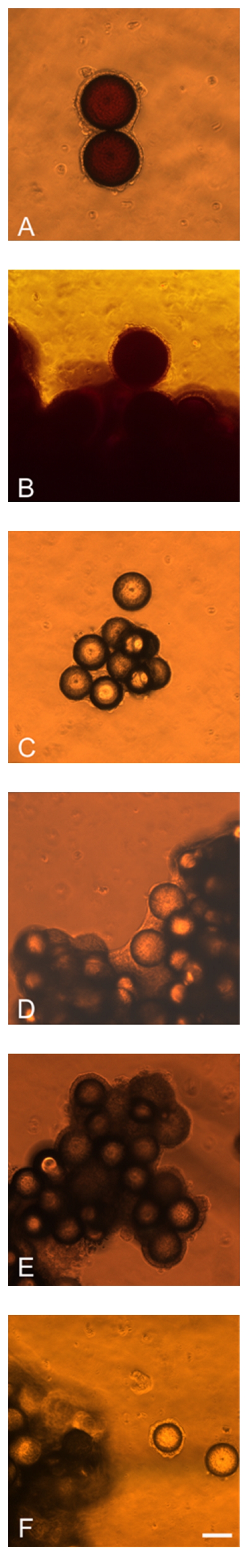
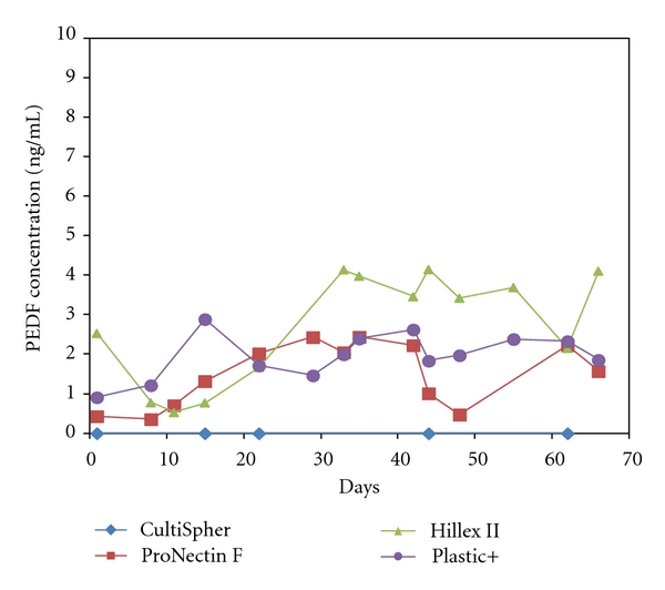
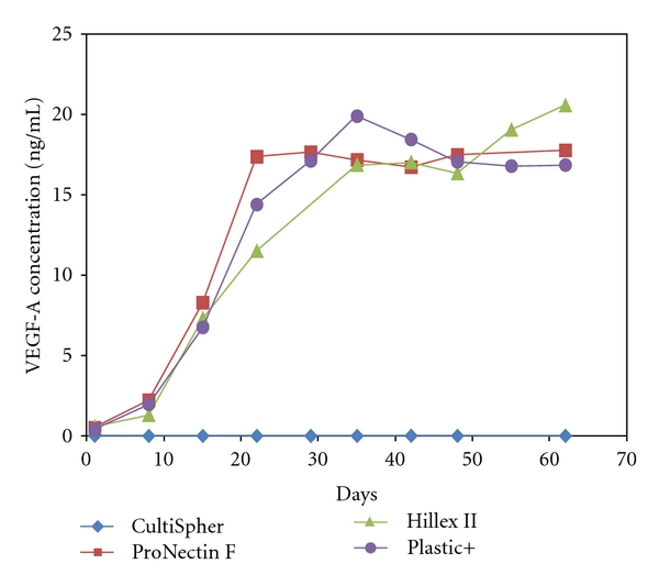
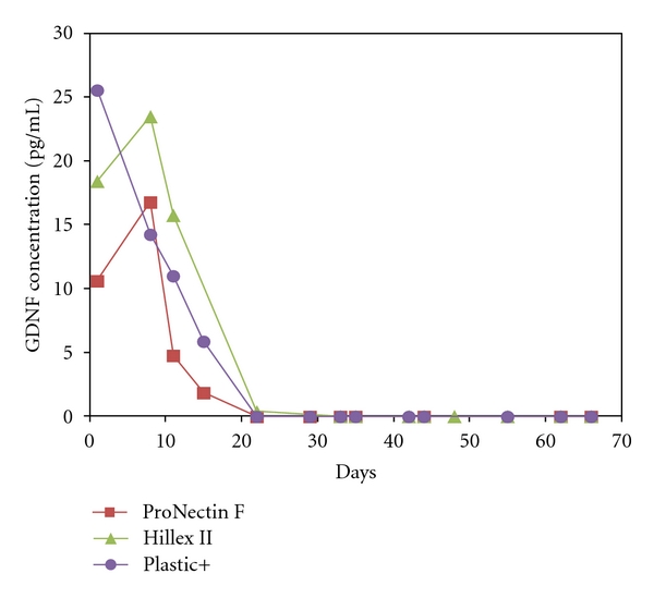
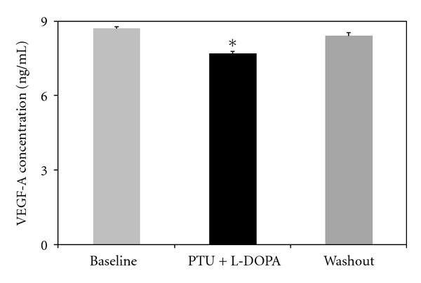
Human retinal pigment epithelial (hRPE) cells have been tested as a cell-based therapy for Parkinson's disease but will require additional study before further clinical trials can be planned. We now show that the long-term survival and neurotrophic potential of hRPE cells can be enhanced by the use of FDA-approved plastic-based microcarriers compared to a gelatin-based microcarrier as used in failed clinical trials. The hRPE cells grown on these plastic-based microcarriers display several important characteristics of hRPE found in vivo: (1) characteristic morphological features, (2) accumulation of melanin pigment, and (3) high levels of production of the neurotrophic factors pigment epithelium-derived factor (PEDF) and vascular endothelial growth factor-A (VEGF-A). Growth of hRPE cells on plastic-based microcarriers led to sustained levels (>1 ng/ml) of PEDF and VEGF-A in conditioned media for two months. We also show that the expression of VEGF-A and PEDF is reciprocally regulated by activation of the GPR143 pathway. GPR143 is activated by L-DOPA (1 μM) which decreased VEGF-A secretion as opposed to the previously reported increase in PEDF secretion. The hRPE microcarriers are therefore novel candidate delivery systems for achieving long-term delivery of the neuroprotective factors PEDF and VEGF-A, which could have a value in neurodegenerative conditions such as Parkinson's disease.


Figures

Figure 1
Figure 1
Example photomicrographs for hRPE cells grown on different plastic-based microcarrier types at 1 month (A, C, E) and two months (B, D, F). Hillex II microcarriers are shown in A and B, Plastic Plus microcarriers are shown in C and D, and ProNectin F microcarriers are illustrated in E and F. Note the characteristic melanin pigment and the polygonal morphology reminiscent of the normal histological features found in vivo. Scale bar = 100 μm.
Figure 2
Figure 2
PEDF concentration time course. The PEDF concentration in the media from the hRPE cells on the different microcarriers was measured with an ELISA. After a short rising phase the PEDF concentration reached a plateau. The hRPE cells on Hillex II, Plastic Plus, and ProNectin F microcarriers secreted comparable amount of PEDF, whereas hRPE cells on the CultiSpher microcarriers did not secrete significant amounts of PEDF.
Figure 3
Figure 3
VEGF-A concentration time course. The VEGF-A concentration in the media from the hRPE cells on the different microcarriers was measured with an ELISA. After a rising phase in the first 20 days after seeding the VEGF-A, concentration reached a plateau and remained stable until the end of the experiment. The hRPE cells on Hillex II, Plastic Plus, and ProNectin F microcarriers secreted comparable amount of VEGF-A, whereas hRPE cells on the CultiSpher microcarriers did not secrete significant amounts of VEGF-A.
Figure 4
Figure 4
GDNF concentration time course. The GDNF concentration in the media from the hRPE cells on the different microcarriers was measured with an ELISA. The hRPE cells on Hillex II, Plastic Plus, and ProNectin F microcarriers secreted comparable amounts of GDNF at the start of the time course. The concentration was an order of magnitude less when compared to VEGF-A and PEDF. The GDNF concentration declined over the first 3 weeks, and there was no detectable amount of GDNF from day 22 on.
Figure 5
Figure 5
Regulation of VEGF-A expression in hRPE cells. Treatment of hRPE monolayers, grown in 24-well plates, with PTU (a tyrosinase blocker used to eliminate endogenous L-DOPA production) and 1 μM L-DOPA leads to a decrease in VEGF-A concentration in the medium. After washout of PTU and L-DOPA, the VEGF-A concentration returns to the baseline levels. Data are presented as the mean of three experiments conducted in triplicate, error bars represent S.E.M., and an asterisk (*) denotes P < 0.0005 using paired t-tests with Bonferroni's correction between the PTU + L-DOPA group from both the baseline and the washout groups.

References

    1. Olanow CW, Stern MB, Sethi K, et al. The scientific and clinical basis for treatment of Parkinson’s disease. Neurology. 2009;72(21, supplement 4):S1–S136. - PubMed
    1. Stocchi F, Olanow CW. Neuroprotection in Parkinson's disease: clinical trials. Annals of Neurology. 2003;53(7, supplement 3):S87–S97. - PubMed
    1. Savitt JM, Dawson VL, Dawson TD. Diagnosis and treatment of Parkinson’s disease: molecules to medicine. Journal of Clinical Investigation. 2006;116(7):1744–1754. - PMC - PubMed
    1. Fahn S, Sulzer D. Neurodegeneration and neuroprotection in Parkinson disease. NeuroRx. 2004;1(1):139–154. - PMC - PubMed
    1. Nutt JG. Motor fluctuations and dyskinesia. In: Factor SA, Weiner WJ, editors. Parkinson's Disease: Diagnosis and Clinical Management. New York, NY, USA: Demos Medical; 2002. pp. 445–453.

Publication types

MeSH terms

LinkOut - more resources