. 2012 Jun 4;9(6):1812-21.
doi: 10.1021/mp3001364. Epub 2012 May 14.

Efficient in vitro siRNA delivery and intramuscular gene silencing using PEG-modified PAMAM dendrimers

Affiliations

Efficient in vitro siRNA delivery and intramuscular gene silencing using PEG-modified PAMAM dendrimers

Yin Tang et al. Mol Pharm. .

Abstract

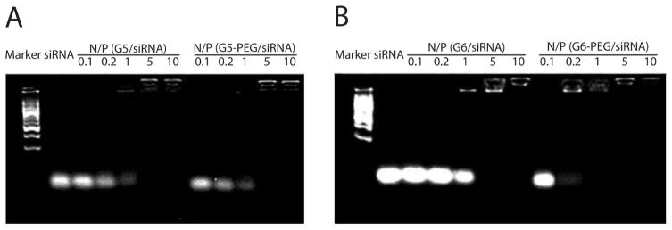
Although siRNA techniques have been broadly applied as a tool for gene knockdown, substantial challenges remain in achieving efficient delivery and in vivo efficacy. In particular, the low efficiency of target gene silencing in vivo is a critical limiting step to the clinical application of siRNA therapies. Poly(amidoamine) (PAMAM) dendrimers are widely used as carriers for drug and gene delivery; however, in vivo siRNA delivery by PAMAM dendrimers remains to be carefully investigated. In this study, the effectiveness of G5 and G6 PAMAM dendrimers with 8% of their surface amines conjugated to MPEG-5000 was studied for siRNA delivery in vitro and for intramuscular in vivo delivery in mice. The results from the PEG-modified dendrimers were compared to the results from the parent dendrimers as well as Lipofectamine 2000 and INTERFERin. Both PEG-modifed dendrimers protect the siRNA from being digested by RNase and gave high transfection efficiency for FITC-labeled siRNA in the primary vascular smooth muscle cells (VSMC) and mouse peritoneal macrophages. The PEG-modified dendrimers achieved knockdown of both plasmid (293A cells) and adenovirus-mediated green fluorescence protein (GFP) expression (Cos7 cells) in vitro with efficiency similar to that shown for Lipofectamine 2000. We further demonstrated in vivo that intramuscular delivery of GFP-siRNA using PEG-modified dendrimer significantly suppressed GFP expression in both transiently adenovirus infected C57BL/6 mice and GFP transgenic mice.


Figures

Fig. 1
Fig. 1
siRNA mobility retardation assays by 3.5% agarose gels. A (from left to right): marker, siRNA alone, G5/siRNA dendriplexes, G5-PEG/siRNA dendriplexes; B (from left to right): marker, siRNA alone, G6/siRNA dendriplexes, G6-PEG/siRNA dendriplexes. The dendriplexes were formed at different N/P ratios from 0.1 to 10. Note that PEGylation does not influence the compacting capability of G5 and G6 dendrimers to siRNA.
Fig. 2
Fig. 2
The protection effect of PEGylated-dendrimers on siRNA in the presence of RNase A. A: siRNA only (control); B: G5-PEG/siRNA dendriplexes formed at an N/P ratio of 10; C: G6-PEG/siRNA dendriplexes formed at an N/P ratio of 10. Note that both G5-PEG and G6-PEG can protect siRNA from being degraded by RNase A at 37 °C for 4h (B & C). However, siRNA alone can be degraded by RNase A within 5 minutes under the same experimental conditions.
Fig. 3
Fig. 3
Effect of PEGylation on the cytotoxicities of G5 (A) and G6 (B) PAMAM dendrimers in Cos-7 cells. Cell viability was determined by CCK-8 assay after incubating the cells with different concentration of dendrimers for 4 hours. Note that the cell viability of Cos-7 cells was significantly increased after PEG-conjugation to G5 and G6 dendrimers. Statistical analysis was done with two-tailed unpaired Student’s t-test (n=3, * p<0.05, *** p<0.005, compared with the parent G5 or G6 at the same concentration).
Fig. 4
Fig. 4
The transfection efficacy of G5- and G6- PEG PAMAM dendrimers to FITC-labeled siRNA in primary VSMC and peritoneal macrophages. Fluorescence images were observed in VSMC (A) and macrophages (B) (left to right: L2000, G5-PEG, G6-PEG.), and fluorescence intensity (C) was measured by flow cytometry. Statistical analysis was done with one-way ANOVA test (n=3, *** p<0.005 vs L2000, ### p<0.005 vs G5-PEG). Note that G5-PEG and G6-PEG have better transfection to FITC-siRNA than L2000 in these two primary cells.
Fig. 5
Fig. 5
Co-transfection of pEGFP and GFP-siRNA to knock down the GFP expression in 293A cells. pEGFP and GFP-siRNA were co-transfected by PEGylated or non- PEGylated G5 or G6 PAMAM dendrimers, and the inhibition efficacy to GFP expression was compared with that of L2000. A: Fluorescence images of co-transfection of pEGFP with non-siRNA (top row) or GFP-siRNA (bottom row). From left to right: L2000, G5, G5-PEG, G6, G6-PEG; B: Fluorescence intensity. Statistical analysis was done with one-way ANOVA test (n=3, * p<0.05, *** p<0.005, compared with the initial GFP expression). Note that G5-PEG has higher co-transfection efficiency and achieves better down-regulation to GFP expression than G6-PEG.
Fig. 6
Fig. 6
Delivery of GFP-siRNA in Cos-7 cells to block down the GFP expression mediated by Ad-GFP. Cos-7 cells were firstly infected by Ad-GFP to express GFP gene, and GFP-siRNA was then transfected by G5-PEG or G6-PEG to block down the GFP expression. A: Fluorescence images of GFP expression in Cos-7 cells. From left to right: top row: Ad-GFP only, Ad-GFP with non-siRNA delivered by L2000, Ad-GFP with GFP-siRNA delivered by L2000; bottom row: Ad-GFP with GFP-siRNA delivered by INTERFERin, Ad-GFP with GFP-siRNA delivered by G5-PEG, Ad-GFP with GFP-siRNA delivered by G6-PEG; B: Relative fluorescence intensity against Ad-GFP. Statistical analysis was done with one-way ANOVA test (n=3, *** p<0.005, compared with non-siRNA transfected by each mediator at the same experimental conditions). Note that compared with L2000 and INTERFERin, G5-PEG and G6-PEG delivered GFP-siRNA achieved the similar down-regulation effects to GFP expression.
Fig. 7
Fig. 7
Delivery of GFP-siRNA to silence the GFP expression mediated by Ad-GFP in C57BL/6 mice. Ad-GFP (1×10⁷ pfu) was injected intramuscularly to the quadriceps of C57BL/6 mice to express GFP gene, and GFP-siRNA with the two doses of 120 and 240 pmol was then delivered respectively to the quadriceps of the mice by G5-PEG or G6-PEG. The gene silence efficacy was investigated at the two time points (48 and 72h) after the GFP-siRNA delivery. Fluorescence images (A) of the muscle’s cryosections of the C57BL/6 mice near the injection point were observed after GFP-siRNA being delivered by G5-PEG (top row) or G6-PEG (bottom row). From left to right: 240 pmol non-siRNA 48h, 120 pmol GFP-siRNA 48h, 240 pmol GFP-siRNA 48h, 120 pmol GFP-siRNA 72h. Relative fluorescence intensity (B) against Ad-GFP infected mice quadriceps was calculated. Statistical analysis was done with one-way ANOVA test (n=5, * p<0.05, ** p<0.01, compared with non-siRNA transfected by each mediator at the same experimental conditions). Note that not only at a high dose (240 pmol) but also at a low dose (120 pmol) of siRNA, G5-PEG delivered GFP-siRNA could accomplish significant down-regulation effect to adenovirus-expressed GFP.
Fig. 8
Fig. 8
Delivery of GFP-siRNA to silence the expression of GFP gene in GFP transgenic mice. GFP-siRNA with the dose of 240 pmol was delivered by G5-PEG or G6-PEG to the quadriceps of the GFP transgenic mice, and the inhibition efficacy to a permanently expressed GFP gene around the injective point was evaluated at the 48 and 72h after the GFP-siRNA delivery. Fluorescence images of the muscle’s cryosections (A) of the GFP transgenic mice near the injection points were observed after GFP-siRNA being delivered by G5-PEG (top row) or G6-PEG (bottom row). From left to right: 240 pmol non-siRNA 48h, 240 pmol GFP-siRNA 48h, 240 pmol GFP-siRNA 72h. Relative fluorescence intensity (B) against the original fluorescence intensity of the GFP mice was calculated. Statistical analysis was done with one-way ANOVA test (n=5, *** p<0.005, compared with non-siRNA transfected by each mediator). Note that G5-PEG delivered GFP-siRNA has significant down-regulation effect to a permanently expressed GFP gene at 48h after siRNA delivery.

References

    1. Bernstein E, Caudy AA, Hammond SM, Hannon GJ. Role for a bidentate ribonuclease in the initiation step of RNA interference. Nature. 2001;409:363–6. - PubMed
    1. Castanotto D, Rossi JJ. The promises and pitfalls of RNA-interference-based therapeutics. Nature. 2009;457:426–33. - PMC - PubMed
    1. Wang J, Lu Z, Wientjes MG, Au JL. Delivery of siRNA therapeutics: barriers and carriers. AAPS J. 2010;12:492–503. - PMC - PubMed
    1. Schiffelers RM, van Rooy I, Storm G. siRNA-mediated inhibition of angiogenesis. Expert Opin Biol Ther. 2005;5:359–68. - PubMed
    1. Olejniczak M, Polak K, Galka-Marciniak P, Krzyzosiak WJ. Recent Advances in Understanding of the Immunological Off-Target Effects of siRNA. Curr Gene Ther. 2011;11:532–43. - PubMed

Publication types