. 2012 Jun 13;31(12):2770-83.
doi: 10.1038/emboj.2012.121. Epub 2012 May 1.

Ubiquitylation of the initiator caspase DREDD is required for innate immune signalling

Affiliations

Ubiquitylation of the initiator caspase DREDD is required for innate immune signalling

Annika Meinander et al. EMBO J. .

Abstract

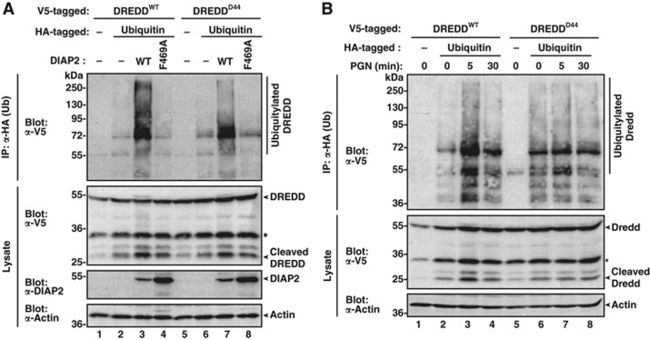
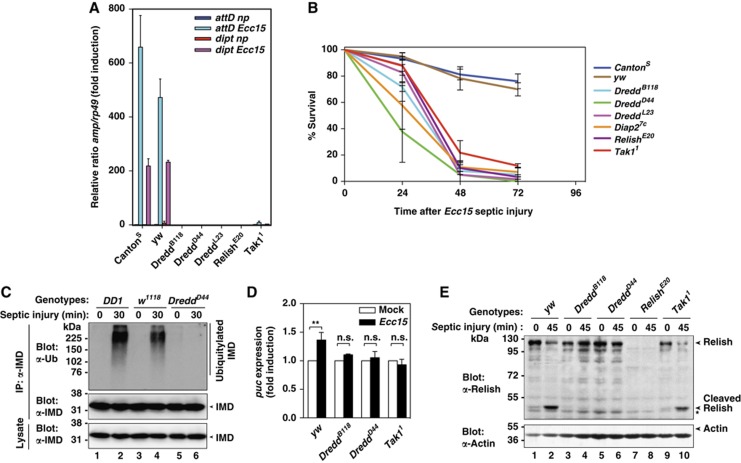
Caspases have been extensively studied as critical initiators and executioners of cell death pathways. However, caspases also take part in non-apoptotic signalling events such as the regulation of innate immunity and activation of nuclear factor-κB (NF-κB). How caspases are activated under these conditions and process a selective set of substrates to allow NF-κB signalling without killing the cell remains largely unknown. Here, we show that stimulation of the Drosophila pattern recognition protein PGRP-LCx induces DIAP2-dependent polyubiquitylation of the initiator caspase DREDD. Signal-dependent ubiquitylation of DREDD is required for full processing of IMD, NF-κB/Relish and expression of antimicrobial peptide genes in response to infection with Gram-negative bacteria. Our results identify a mechanism that positively controls NF-κB signalling via ubiquitin-mediated activation of DREDD. The direct involvement of ubiquitylation in caspase activation represents a novel mechanism for non-apoptotic caspase-mediated signalling.


Conflict of interest statement

The authors declare that they have no conflict of interest.

Figures

Figure 1
Figure 1
DIAP2 binds and ubiquitylates the initiator caspase DREDD. (A, B) DIAP2’s RING finger and both BIR domains are required to resist Ecc15 bacterial infection. Diap27c mutant animals were reconstituted with rescue constructs expressing the indicated DIAP2 proteins. To confirm the requirement of DIAP2 E3 ligase activity for the IMD pathway (A), DIAP2 constructs were ubiquitously expressed in transgenic flies under the control of daughterless (Da)-GAL4. Animals were pricked with a needle previously dipped into Ecc15 and the survival rates monitored. To evaluate the contribution of individual BIR domain of DIAP2 (B), transgenic flies were generated using germ-line-specific phiC31 integration (Bischof et al, 2007). Transgenes were expressed using the fat-body-specific c564-Gal4 driver line. Animals were injected with 13.8 nl of Ecc15 bacterial solution (optical density of 300) and the survival rates monitored. The experiments were performed using at least 20 flies for each genotype. Shown is a representative experiment of at least three repeats. C466Y represents a RING mutant of DIAP2, which lacks E3 ligase activity (Ribeiro et al, 2007), D163 and D263A disrupts the BIR pocket of BIR2 and BIR3, respectively. (C, D) DIAP2 binds to DREDD. Reciprocal binding assays with DIAP2 and DREDD. The indicated constructs were transfected into S2 cells and GST-tagged DIAP2 (C) and V5-tagged DREDD (D) were purified with Glutathione resin and α-V5 antibodies, respectively. The expression and the presence of the co-purified proteins were assessed by immunoblotting with the indicated antibodies. (E) DIAP2 ubiquitylates DREDD in a RING finger-dependent manner. HA–Ub was co-expressed with DREDD–V5 in S2 cells together with vector control, DIAP2WT or the DIAP2 RING mutants DIAP2C466Y and DIAP2F469A. Cells were lysed under denaturing conditions and ubiquitylated proteins isolated using α-HA antibodies. The presence and ubiquitylation of DREDD and DIAP2 was assessed with the indicated antibodies. (F) DREDD is transiently ubiquitylated in a signal-dependent manner. S2 cells were transfected with DREDD–V5. Subsequently, cells were treated with 1 μg/ml DAP-PGN for the indicated time points before being harvested. DREDD was immunoprecipitated using α-V5 resin and ubiquitylated DREDD analysed using α-Ub antibodies. (G) DIAP2 promotes the conjugation of K63-linked Ub chains on DREDD. The indicated constructs were transfected into S2 cells, and HA-tagged DREDD and IMD were purified under denaturing conditions. The presence of the purified proteins and identity of the respective Ub chains were detected by immunoblotting using the indicated antibodies (see Materials and methods for details).
Figure 2
Figure 2
DIAP2 binds to the DED1 of DREDD. (A) Schematic representation of the DIAP2 and DREDD constructs used in this study. (B) DIAP2 binds to the pro-domain of DREDD. Co-IP assays with DIAP2 and various fragments of DREDD. S2 cells were co-transfected with HA-tagged DIAP2 and V5-tagged DREDD. Expression and the presence of co-purified proteins were analysed by immunoblotting with the indicated antibodies. (C) DIAP2 associates with DED1 of DREDD. Co-IPs were performed with DIAP2 and individual DEDs or the prodomain of DREDD as in (B). (D) dFADD also binds to the DED1 of DREDD. Co-IPs were performed with dFADD and individual DEDs or the prodomain of DREDD as in (B). (E) Competition assay with DIAP2, dFADD and DREDD indicates that DIAP2 and dFADD can bind to DREDD simultaneously. Co-IPs were performed with dFADD, DIAP2 and DREDD. Expression and the presence of co-purified proteins were analysed as in (B). (F) The N-terminal portion of DIAP2 interacts with DREDD. Co-IPs were performed with DREDD and the indicated fragments of DIAP2. Expression and the presence of co-purified proteins were analysed as in (B). (G) The BIR2 and BIR3 domains mediate binding to DREDD. Co-IPs were performed with DREDD and individual BIR domains of DIAP2. Expression and the presence of co-purified proteins were analysed as in (B). An asterisk marks a cleavage product of BIR3. (H) The interaction between DIAP2 and DREDD does not require the hydrophobic pocket of the BIR2 and BIR3. Co-IPs were performed with DREDD and the indicated WT or mutant BIR domains. Expression and the presence of co-purified proteins were analysed as in (B). (I) In contrast, the binding of DIAP2 to cleaved IMD critically depends on the hydrophobic pockets of the BIR2 and BIR3. IMD corresponds to the DREDD-cleaved form of IMD (aa 31–273) and was expressed using the Ub-fusion technique (Varshavsky, 2000; Tenev et al, 2005). Co-IPs with DREDD and the indicated WT or mutant BIR domains were performed as in (H). BIR1/2/3 encompasses DIAP2’s N-terminal segment that carries the BIR1, BIR2 and BIR3 domains.
Figure 3
Figure 3
The G120R loss-of-function mutation of DREDD disrupts DIAP2-mediated ubiquitylation. (A) Schematic diagram of the domain architecture of DREDD. Residue G120 maps to the DED1 of DREDD. (B) Ribbon representation of the prodomain structure of DREDD (residues 40–235). The 3D structure of DREDD’s prodomain was modelled using the X-ray structure of the DEDs of the viral-FLIP MC159 (pdb 2bbz) as a template. The six-helical bundle structure of the death fold of DED1 is indicated. The position of G120 is highlighted in red. (C) Molecular surfaces of the same residues as shown in (B). G120 and G120R of WT DREDD (left panel) and DREDDD44 (right panel) are highlighted in red. DED1 is indicated in green, while the DED2 is depicted in grey. (D) DREDDD44 binds to DIAP2 like WT DREDD. Co-IPs were performed with DIAP2 and DREDDWT and DREDDD44. Expression and the presence of co-purified proteins were analysed by immunoblotting with the indicated antibodies. Of note, DIAP2 only co-purifies full-length DREDD, but not processed DREDD (p20-10) that lacks the pro-domain. (E) The G120R mutation does not affect the ability of DREDD to bind to dFADD. Co-IPs with dFADD and DREDDWT or DREDDD44 were performed as in (D). (F, G) DREDDD44 is not impaired in its ability to hetero- or homodimerize with WT and mutant DREDDD44. Co-IPs with DREDDWT and DREDDD44 were performed as in (D). (H) The G120R mutation does not affect the inherent catalytic activity of DREDD, since DREDDD44 can cleave Relish similar to DREDDWT upon overexpression. S2 cells were co-transfected with FLAG-tagged Relish and empty vector, V5-tagged WT, catalytically inactive (C408A) or G120R mutant DREDD. Relish cleavage was assessed by immunoblotting using α-FLAG antibodies. (I) The G120R mutation does not affect DREDD’s protein stability. Cycloheximide (CHX) chase experiments to compare the protein stability of DREDDWT and DREDDD44. Equal loading was assessed with α-actin antibodies.
Figure 4
Figure 4
The G120R mutation impairs DREDD ubiquitylation. (A) DIAP2 ubiquitylates DREDDD44 less efficiently than WT DREDD. HA–Ub was co-expressed with DREDDWT and DREDDD44 in S2 cells together with vector control, DIAP2WT or the DIAP2 RING mutant DIAP2F496A. Cells were lysed under denaturing conditions and ubiquitylated proteins isolated using α-HA antibodies. The presence of DREDD and DIAP2 and the DREDD ubiquitylation was assessed with the indicated antibodies. (B) DREDDD44 fails to be ubiquitylated in a signal-dependent manner. S2 cells were co-transfected with HA–Ub and DREDDWT or DREDDD44. Subsequently, cells were treated with 1 μg/ml DAP-PGN for the indicated time points before being harvested under denaturing conditions. Ubiquitylated DREDD was purified and detected as described in (A). An asterisk marks a cross-reactive band.
Figure 5
Figure 5
The G120R mutation impairs signal-dependent cleavage of Relish and Imd, and abrogates activation of AMP gene expression. (A) Quantitative RT–PCR analysis of attacin D and diptericin induction in the indicated fly strains. rp49 was used as the experimental expression standard. Shown are the relative expression ratios of attacin D/rp49 and diptericin/rp49 5 h post-infection. Values are normalized to non-pricked CantonS. Np stands for non-pricked. Error bars indicate SEM from three independent experimental repeats using at least 20 flies per repeat. (B) DreddD44 mutant flies die upon infection with Ecc15. Adult flies of the indicated genotypes were subjected to Ecc15-mediated septic injury and their survival rates were monitored at the indicated time points. Error bars indicate SEM from three independent experimental repeats using at least 20 flies per repeat. (C) Immunoblot analysis assessing ubiquitylation of IMD, which requires DREDD-mediated cleavage of IMD (Paquette et al, 2010). The indicated fly strains were exposed to septic injury and fly lysates were used to immunoprecipitate IMD. The presence and modification of IMD was subsequently analysed by immunoblotting with the indicated antibodies. DD1 and w1118 fly strains are WT strains that served as controls. (D) IMD-mediated activation of JNK in the indicated fly strains was measured by evaluating expression of puckered, a bona fide downstream target of the JNK signal transduction cascade. Values were normalized to mock injections. The asterisks indicate a P-value of P<0.01, calculated on raw data before normalization. Ns designates non-significant differences. (E) Immunoblot analysis assessing cleavage of Relish in the indicated fly strains. While Relish cleavage occurred in a signal-dependent manner in yw flies, Relish failed to be cleaved in DreddB118 and DreddD44 following septic injury with Ecc15. In contrast, Relish was partially cleaved in Tak11 mutant flies. Equal loading was assessed with α-actin antibodies. An asterisk indicates a cross-reactive band.
Figure 6
Figure 6
Model depicting Ub-dependent activation of NF-κB during IMD signalling. (1) Ligand binding induces recruitment of IMD, DREDD and dFADD to PGRP-LCx. (2) Recruitment of DIAP2 targets DREDD for K63-linked ubiquitylation. (3) This allows Ub-mediated aggregation, activation and processing of DIAP2-modified DREDD. (4) Active DREDD subsequently cleaves IMD, which exposes an IBM at the neo-amino-terminus of cleaved IMD. The IBM of IMD subsequently binds to the BIR2/3 of DIAP2. (5) This provides DIAP2 with an additional contact point within the signalling complex, reinforcing complex stability and permitting DIAP2-mediated ubiquitylation of IMD, and quite possibly other components of the signalling complex. (6) The Ub chains on IMD and DREDD may serve as scaffolds for the recruitment of dTAK1 and IKK. Since IKK can bind to Relish, as evidenced by its ability to phosphorylate Relish, IKK might also bring Relish into close proximity of ubiquitylated and active DREDD, allowing DREDD-mediated proteolysis of Relish. (7) The proximity to the signalling complex also allows phospho-mediated activation of Relish. (8) Subsequently, modified Relish translocates to the nucleus where (9) it drives expression of AMP target genes.

Comment in

References

    1. Bischof J, Maeda RK, Hediger M, Karch F, Basler K (2007) An optimized transgenesis system for Drosophila using germ-line-specific phiC31 integrases. Proc Natl Acad Sci USA 104: 3312–3317 - PMC - PubMed
    1. Chen P, Rodriguez A, Erskine R, Thach T, Abrams JM (1998) Dredd, a novel effector of the apoptosis activators reaper, grim, and hid in Drosophila. Dev Biol 201: 202–216 - PubMed
    1. Choe KM, Lee H, Anderson KV (2005) Drosophila peptidoglycan recognition protein LC (PGRP-LC) acts as a signal-transducing innate immune receptor. Proc Natl Acad Sci USA 102: 1122–1126 - PMC - PubMed
    1. Choe KM, Werner T, Stoven S, Hultmark D, Anderson KV (2002) Requirement for a peptidoglycan recognition protein (PGRP) in Relish activation and antibacterial immune responses in Drosophila. Science 296: 359–362 - PubMed
    1. Chun HJ, Zheng L, Ahmad M, Wang J, Speirs CK, Siegel RM, Dale JK, Puck J, Davis J, Hall CG, Skoda-Smith S, Atkinson TP, Straus SE, Lenardo MJ (2002) Pleiotropic defects in lymphocyte activation caused by caspase-8 mutations lead to human immunodeficiency. Nature 419: 395–399 - PubMed

Publication types

MeSH terms