. 2012 Aug;32(8):1589-99.
doi: 10.1038/jcbfm.2012.58. Epub 2012 May 2.

Effect of mild cognitive impairment and APOE genotype on resting cerebral blood flow and its association with cognition

Affiliations

Effect of mild cognitive impairment and APOE genotype on resting cerebral blood flow and its association with cognition

Christina E Wierenga et al. J Cereb Blood Flow Metab. 2012 Aug.

Abstract

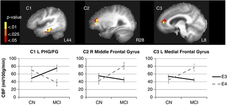
Using whole-brain pulsed arterial spin labeling magnetic resonance imaging, resting cerebral blood flow (CBF) was measured in 20 mild cognitive impairment (MCI; 11 ɛ3 and 9 ɛ4) and 40 demographically matched cognitively normal (CN; 27 ɛ3 and 13 ɛ4) participants. An interaction of apolipoprotein (APOE) genotype (ɛ3 and ɛ4) and cognitive status (CN and MCI) on quantified gray-matter CBF corrected for partial volume effects was found in the left parahippocampal and fusiform gyri (PHG/FG), right middle frontal gyrus, and left medial frontal gyrus. In the PHG/FG, CBF was elevated for CN ɛ4 carriers but decreased for MCI ɛ4 carriers. The opposite pattern was seen in frontal regions: CBF was decreased for CN ɛ4 carriers but increased for MCI ɛ4 carriers. Cerebral blood flow in the PHG/FG was positively correlated with verbal memory for CN ɛ4 adults (r=0.67, P=0.01). Cerebral blood flow in the left medial frontal gyrus was positively correlated with verbal memory for MCI ɛ4 adults (r=0.70, P=0.05). Findings support dynamic pathophysiologic processes in the brain associated with Alzheimer's disease risk and indicate that cognitive status and APOE genotype have interactive effects on CBF. Correlations between CBF and verbal memory suggest a differential neurovascular compensatory response in posterior and anterior cortices with cognitive decline in ɛ4 adults.


Figures

Figure 1
Figure 1
The interaction of cognitive status (CN and MCI) and APOE genotype (ɛ3 and ɛ4) on cerebral blood flow (CBF). Thresholded and clustered results (protecting a whole-brain voxelwise P<0.05; red: P<0.05, orange: P<0.025, yellow: P<0.01) for a two-way analysis of variance indicating an interaction of cognitive status (CN and MCI) and APOE genotype (ɛ3 and ɛ4) with corresponding graphical presentation of significant CBF differences. Error bars represent the standard error of the mean. Results are overlaid onto sagittal slices of a high-resolution anatomical image averaged across all participants (L: left; R: right; C: cluster; PHG/FG: parahippocampal gyrus/fusiform gyrus). For the interaction of cognitive status by APOE genotype: C1: CN ɛ3=46.0±17.0, CN ɛ4=70.3±45.5, MCI ɛ3=75.6±43, MCI ɛ4=37.7±12.7, F(3,56)=4.5, P<0.01; C2: CN ɛ3=64.1±31.5, CN ɛ4=42.2±18.8, MCI ɛ3=45.0±17.4, MCI ɛ4=80.9±33.6, F(3,56)=4.8, P<0.01; C3: CN ɛ3=58.9±23.2, CN ɛ4=44.8±11.8, MCI ɛ3=44.6±14.6, MCI ɛ4=76.8±40.3, F(3,56)=4.4, P<0.01. APOE, apolipoprotein; CN, cognitively normal; MCI, mild cognitive impairment.
Figure 2
Figure 2
The effect of cognitive status on cerebral blood flow (CBF). Thresholded and clustered results (protecting a whole-brain voxelwise P<0.05; red: P<0.05, orange: P<0.025, yellow: P<0.01) for a two-way analysis of variance indicating a main effect of cognitive status (bottom right) with corresponding graphical presentation of significant CBF differences. Error bars represent the standard error of the mean. Results are overlaid onto axial slices of a high-resolution anatomical image averaged across all participants (L: left; R: right; I: inferior; S: superior; c: cluster; STG: superior temporal gyrus; Hipp: hippocampus). For main effect of cognitive status: C1: CN=20.9±8.7, MCI=41.6±18.5, F(1,58)=34.1, P<0.001; C2: CN=29.5±10.9, MCI=53.2±25.3, F(1,58)=25.4, P<0.001; C3: CN=32.0±13.6, MCI=52.5±20.3, F(1,58)=21.4, P<0.001; C4: CN=41.7±15.7, MCI=67.2±31.0, F(1,58)=17.8, P<0.001; C5: CN=29.9±12.0, MCI=48.2±20.0, F(1,58)=19.2, P<0.001. CN, cognitively normal; MCI, mild cognitive impairment; SFG, superior frontal gyrus.

References

    1. Alsop DC, Detre JA, Grossman M. Assessment of cerebral blood flow in Alzheimer's disease by spin-labeled magnetic resonance imaging. Ann Neurol. 2000;47:93–100. - PubMed
    1. Braak H, Braak E. Neuropathological staging of Alzheimer-related changes. Acta Neuropathol (Berl) 1991;82:239–259. - PubMed
    1. Brown GG, Clark C, Liu TT. Measurement of cerebral perfusion with arterial spin labeling: Part 2. Applications. J Int Neuropsychol Soc. 2007;13:526–538. - PMC - PubMed
    1. Brown GG, Eyler Zorrilla LT, Georgy B, Kindermann SS, Wong EC, Buxton RB. BOLD and perfusion response to finger-thumb apposition after acetazolamide administration: differential relationship to global perfusion. J Cereb Blood Flow Metab. 2003;23:829–837. - PubMed
    1. Brumm KP, Perthen JE, Liu TT, Haist F, Ayalon L, Love T. An arterial spin labeling investigation of cerebral blood flow deficits in chronic stroke survivors. Neuroimage. 2010;51:995–1005. - PMC - PubMed

Publication types

Substances