Review
. 2012 Jul;32(7):1393-415.
doi: 10.1038/jcbfm.2012.53. Epub 2012 May 2.

Noninvasive molecular imaging of neuroinflammation

Collaborators, Affiliations
Review

Noninvasive molecular imaging of neuroinflammation

Andreas H Jacobs et al. J Cereb Blood Flow Metab. 2012 Jul.

Abstract

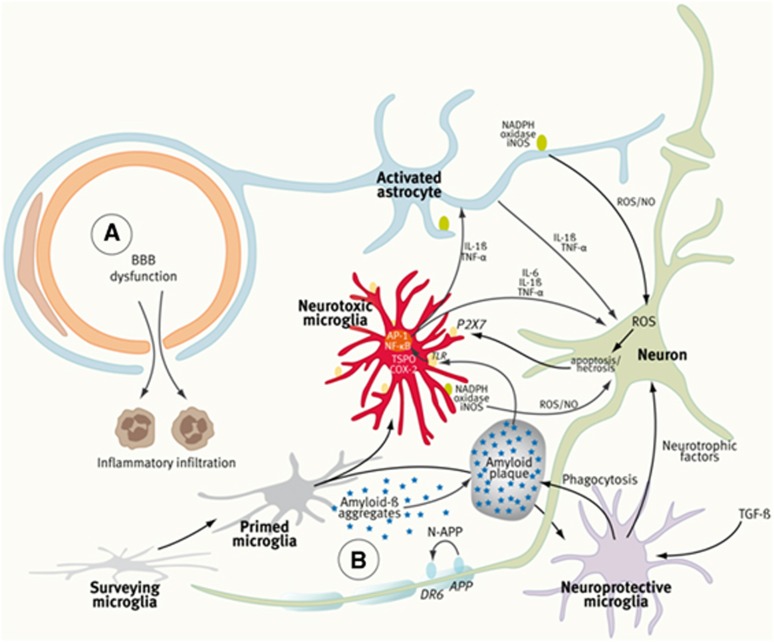
Inflammation is a highly dynamic and complex adaptive process to preserve and restore tissue homeostasis. Originally viewed as an immune-privileged organ, the central nervous system (CNS) is now recognized to have a constant interplay with the innate and the adaptive immune systems, where resident microglia and infiltrating immune cells from the periphery have important roles. Common diseases of the CNS, such as stroke, multiple sclerosis (MS), and neurodegeneration, elicit a neuroinflammatory response with the goal to limit the extent of the disease and to support repair and regeneration. However, various disease mechanisms lead to neuroinflammation (NI) contributing to the disease process itself. Molecular imaging is the method of choice to try to decipher key aspects of the dynamic interplay of various inducers, sensors, transducers, and effectors of the orchestrated inflammatory response in vivo in animal models and patients. Here, we review the basic principles of NI with emphasis on microglia and common neurologic disease mechanisms, the molecular targets which are being used and explored for imaging, and molecular imaging of NI in frequent neurologic diseases, such as stroke, MS, neurodegeneration, epilepsy, encephalitis, and gliomas.
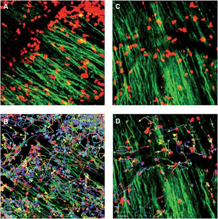

Figures

Figure 1
Figure 1
Components of the neurovascular unit (NVU), the ‘Vicious Cycle' of neuroinflammation, and the ‘Ying and Yang' of microglia. In ischemic stroke and multiple sclerosis (MS) (A), peripheral immune cells contribute substantially to the local inflammatory response and tissue damage. The dynamics of blood–brain barrier (BBB) penetration of immune cells is increasingly being studied in vivo by two-photon microscopy at the cellular level and by ultrasmall superparamagnetic iron oxides (USPIOs) (phagocytosed by peripheral macrophages) and magnetic resonance imaging (MRI). In neurodegenerative diseases (B), the BBB is intact and neuroinflammation (NI) is predominated by the activation of microglia cells, which is mostly studied by translocator protein (TSPO)-targeting tracers and positron-emission tomography (PET) or single-photon emission computed tomography (SPECT) imaging. In Alzheimer's disease (AD), for example, Aß forms aggregates that activate microglia through Toll-like receptor (TLR) and receptor for advanced glycation end products (RAGE). Activated microglia secrete inflammatory mediators such as interleukin (IL)-6, IL-1β, and tumor necrosis factor (TNF)-α to coactivate astrocytes and to induce neuronal death, which in turn will amplify microglia activation through purinergic P2X7 receptors. Protective microglia mediate Aβ clearance, removal of cell debris, and promote neuroregeneration. It should be stated that the clear distinction between ‘surveying,' ‘primed,' and ‘activated' microglia may be an oversimplification of the complex molecular guidance of various microglia functional states. (Figure prepared according to Ferrari and Tarelli, 2011; Glass et al, 2010; and Moskowitz et al, 2010.) COX-2, cyclooxygenase-2; AP-1, activator protein; NF-κB, nuclear factor-κB; iNOS, nitric oxide synthase; ROS, reactive oxygen species; NO, nitric oxide; TGF-β, tumor growth factor-β.
Figure 2
Figure 2
Time course of microglia activation after experimental ischemia as determined by [18F]DPA-714 and μPET. Panels (AG) depict images of different rats showing [18F]DPA-714 accumulation in the ischemic hemisphere at various time points (1 to 30 days after stroke). Panels (H, I) depict quantitative [18F]DPA-714 accumulation in ischemic (H) versus control (I) hemisphere showing the peak intensities of microglia activation in the 7 to 15 day after stroke period. (Figure reprinted with permission from Martin et al, 2010.) ID, injected dose; PET, positron-emission tomography.
Figure 3
Figure 3
Location and extent of microglia activation after focal subcortical ischemia as determined by [11C](R)-PK11195 and HRRT-PET in conjunction with diffusion tensor imaging (DTI) MRI. Various microglia activation patterns can be observed after focal cerebral ischemia in humans depending on the affection of the pyramidal tract and the primary lesion size. Acutely activated microglia at the infarct site that decreases after 6 months (A) are related with good clinical outcome although microglia activation in the brain stem as sign of anterograde axonal degeneration persists. Lesions that cause a complete transection of the pyramidal tract as determined by DTI are related to persistent microglia activity at the site of infarction and in the brain stem at 6 months and are related to a poor outcome (B). In patients where the pyramidal tract is not affected, microglia activation occurs only at the infarct site and not in the brain stem (C). (Figure reprinted with permission from Thiel et al, 2010.) FA, fractional anisotropy; HRRT, high resolution research tomograph; MRI, magnetic resonance imaging; PET, positron-emission tomography.
Figure 4
Figure 4
Cross-sectional patterns of lesion enhancement in patients with multiple sclerosis (MS) as detected by magnetic resonance imaging (MRI). (A) Pre-Gd T2SE images showing MS lesions. (B) Pre-GdT1-w images showing hypointensity of some of the MS lesions. (C) Some lesions are Gd-DTPA positive. (D) Post-USPIO images show a Gd-DTPA-positive/USPIO ring-enhancing lesion (arrow) and a Gd-DTPA-negative/focally USPIO-positive lesion (arrowhead). (Figure reprinted with permission from Vellinga et al, 2008.) USPIO, ultrasmall superparamagnetic iron oxide; Gd-DTPA, gadolinium-diethylenetriaminepentaacetic acid.
Figure 5
Figure 5
Immune infiltrates in demyelinating lesions as depicted by two-photon laser scanning microscopy (TPLSM) are highly dynamic and show different motility patterns in distinct disease stages. (A) TPLSM of a representative brainstem lesion in the onset of EAE in B6.tdRFP/B6.Thy1.EGFP (green: EGFP, neuronal processes; red: CD45+.tdRFP cells) (maximal intensity projection of a volume of 70 mm thickness and 36 planes, time point 0). (B) Automated single-cell tracking of CD45+.tdRFP cells. (C) TPLSM of a representative brainstem lesion in the onset of 2d2.tdRFP Th17 cells (red)-induced passive EAE in B6.Rag1/Thy1.EGFP. (D) Automated single-cell tracking of 2d2.tdRFP Th17 cells. (Figure reprinted with permission from Siffrin et al, 2010).
Figure 6
Figure 6
Early detection of amyloid-β as damage-associated molecular pattern (DAMP) and microglia activation in patients with mild cognitive impairment by positron-emission tomography (PET). [11C]-(R)-PK11195 binding potentials (BPs) and corresponding [11C]PIB ratio images in two patients with PIB-positive mild cognitive impairment (MCI). MCI-I has normal PK binding and MCI-II has increased PK binding suggesting that Aβ deposition may be related but also unrelated to microglia activation at very early disease stages. The detection of microglial activation in patients with MCI suggests that anti-inflammatory therapies may be relevant to the prevention of AD. (Figure reprinted with permission from Okello et al, 2009.)

References

    1. Abourbeh G, Thézé B, Maroy R, Dubois A, Dollé F, Tavitian B, Boisgard R.2012Imaging microglial/macrophage activation in spinal cords of experimental autoimmune encephalomyelitis (EAE) rats by PET using the mitochondrial 18-kda translocator protein (TSPO) radioligand, [18F]-DPA-714 J Neurosci(in press) - PMC - PubMed
    1. Ajami B, Bennett JL, Krieger C, McNagny KM, Rossi FM. Infiltrating monocytes trigger EAE progression, but do not contribute to the resident microglia pool. Nat Neurosci. 2011;14:1142–1149. - PubMed
    1. Alexopoulos GS, Morimoto SS.2011The inflammation hypothesis in geriatric depression Int J Geriatr Psychiatryadvance online publication 2 March 2011; doi:10.1002/gps.2672(e-pub ahead of print) - DOI - PMC - PubMed
    1. Allan SM, Tyrrell PJ, Rothwell NJ. Interleukin-1 and neuronal injury. Nat Rev Immunol. 2005;5:629–640. - PubMed
    1. Amor S, Puentes F, Baker D, van der Valk P. Inflammation in neurodegenerative diseases. Immunology. 2010;129:154–169. - PMC - PubMed