

Oral pigmented lesions: Clinicopathologic features and review of the literature
- PMID: 22549672
- PMCID: PMC3505710
- DOI: 10.4317/medoral.17679
Oral pigmented lesions: Clinicopathologic features and review of the literature
Abstract
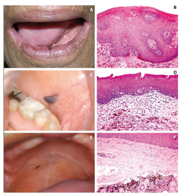
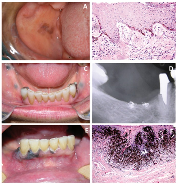
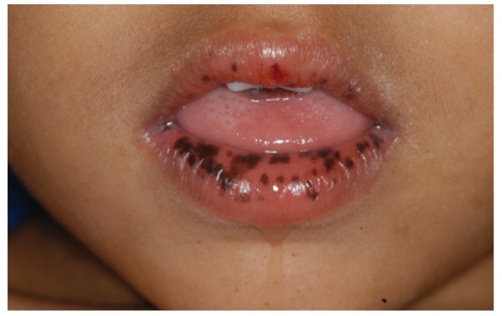
Diagnosis of pigmented lesions of the oral cavity and perioral tissues is challenging. Even though epidemiology may be of some help in orientating the clinician and even though some lesions may confidently be diagnosed on clinical grounds alone, the definitive diagnosis usually requires histopathologic evaluation. Oral pigmentation can be physiological or pathological, and exogenous or endogenous. Color, location, distribution, and duration as well as drugs use, family history, and change in pattern are important for the differential diagnosis. Dark or black pigmented lesions can be focal, multifocal or diffuse macules, including entities such as racial pigmentation, melanotic macule, melanocytic nevus, blue nevus, smoker's melanosis, oral melanoacanthoma, pigmentation by foreign bodies or induced by drugs, Peutz-Jeghers syndrome, Addison's disease and oral melanoma. The aim of this review is to present the main oral black lesions contributing to better approach of the patients.
Figures
References
-
- Kauzman A, Pavone M, Blanas N, Bradley G. Pigmented lesions of the oral cavity: review, differential diagnosis, and case presentation. J Can Dent Assoc. 2004;70:682–3. - PubMed
-
- Meleti M, Vescovi P, Mooi WJ, van der Waal I. Pigmented lesions of the oral mucosa and perioral tissues: a flow-chart for the diagnosis and some recommendations for the management. Oral Surg Oral Med Oral Pathol Oral Radiol Endod. 2008;105:606–16. - PubMed
-
- Eisen D. Disorders of pigmentation in the oral cavity. Clin Dermatol. 2000;18:579–87. - PubMed
-
- Ishikawa I, Aoki A, Takasaki A. Potential applications of Erbium: YAG laser in periodontics. J Periodont Res. 2004;39:275–85. - PubMed
-
- Halder RM, Nootheti PK. Ethnic skin disorders overview. J Am Acad Dermatol. 2003;48:S143–8. - PubMed
Publication types
MeSH terms
LinkOut - more resources
Full Text Sources
Medical
