

Osseous reaction to implantation of two endodontic cements: Mineral trioxide aggregate (MTA) and calcium enriched mixture (CEM)
- PMID: 22549692
- PMCID: PMC3482541
- DOI: 10.4317/medoral.18136
Osseous reaction to implantation of two endodontic cements: Mineral trioxide aggregate (MTA) and calcium enriched mixture (CEM)
Abstract
Aim: The aim of the present in vivo study was to determine bone tissue reaction to calcium enriched mixture (CEM) and mineral trioxide aggregate (MTA) using a rat femur model.
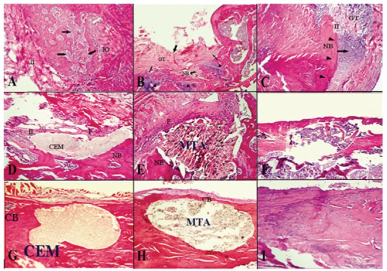
Study design: Sixty-three rats were selected and randomly divided into three groups of 21 each [experimental groups (n=15), control (n=6)]. Implantation cavities were prepared in each femoral bone and randomly filled with the biomaterials only in the experimental groups. The animals in three groups were sacrificed 1, 4, and 8 weeks postoperatively. Histologic evaluations comprising inflammation severity and new bone formation were blindly made on H&E-stained decalcified 6-µm sections.
Results: At 1, 4, and 8 weeks after implantation number of inflammatory cells had decreased in the CEM, MTA and control groups, respectively, with no statistically significant differences. Conversely, new bone formation had increased in all the experimental and control groups, without statistically significant differences.
Conclusion: The results suggest that biocompatibility of MTA, as gold standard, and CEM cement as a new endodontic biomaterial are comparable.
Figures
References
-
- Torabinejad M, Watson TF, Pitt-Ford TR. Sealing ability of a mineral trioxide aggregate when used as a root end filling material. J Endod. 1993 ;19:591–5. - PubMed
-
- Funteas UR, Wallace JA, Fochtman EW. A comparative analysis of mineral trioxide aggregate and Portland cement. Aust Endod J. 2003;29:43–4. - PubMed
-
- Shahi S, Rahimi S, Lotfi M, Yavari HR, Gaderian AR. A comparative study of biocompatibility of three root-end filing materials in rat connective tissue. J Endod. 2006;32:776–80. - PubMed
-
- Lotfi M, Vosoughhosseini S, Saghiri MA, Mesgariabbasi M, Ranjkesh B. Effect of white mineral trioxide aggregate mixed with disodium hydrogen phosphate on inflammatory cells. J Endod. 2009;35:703–5. - PubMed
-
- Rahimi S, Shahi S, Lotfi M, Yavari HR, Charejoo ME. Comparison of microleakage with three different thicknesses of mineral trioxide aggregate as root-end filling material. J Oral Sci. 2008;50:273–7. - PubMed
MeSH terms
Substances
LinkOut - more resources
Full Text Sources
