. 2012 Jun 15;287(25):20876-87.
doi: 10.1074/jbc.M112.344929. Epub 2012 May 1.

Increased resting intracellular calcium modulates NF-κB-dependent inducible nitric-oxide synthase gene expression in dystrophic mdx skeletal myotubes

Affiliations

Increased resting intracellular calcium modulates NF-κB-dependent inducible nitric-oxide synthase gene expression in dystrophic mdx skeletal myotubes

Francisco Altamirano et al. J Biol Chem. .

Abstract

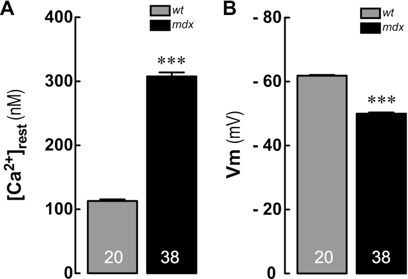
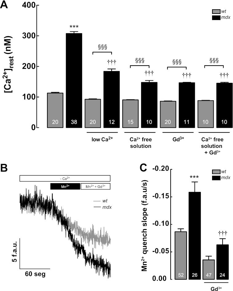
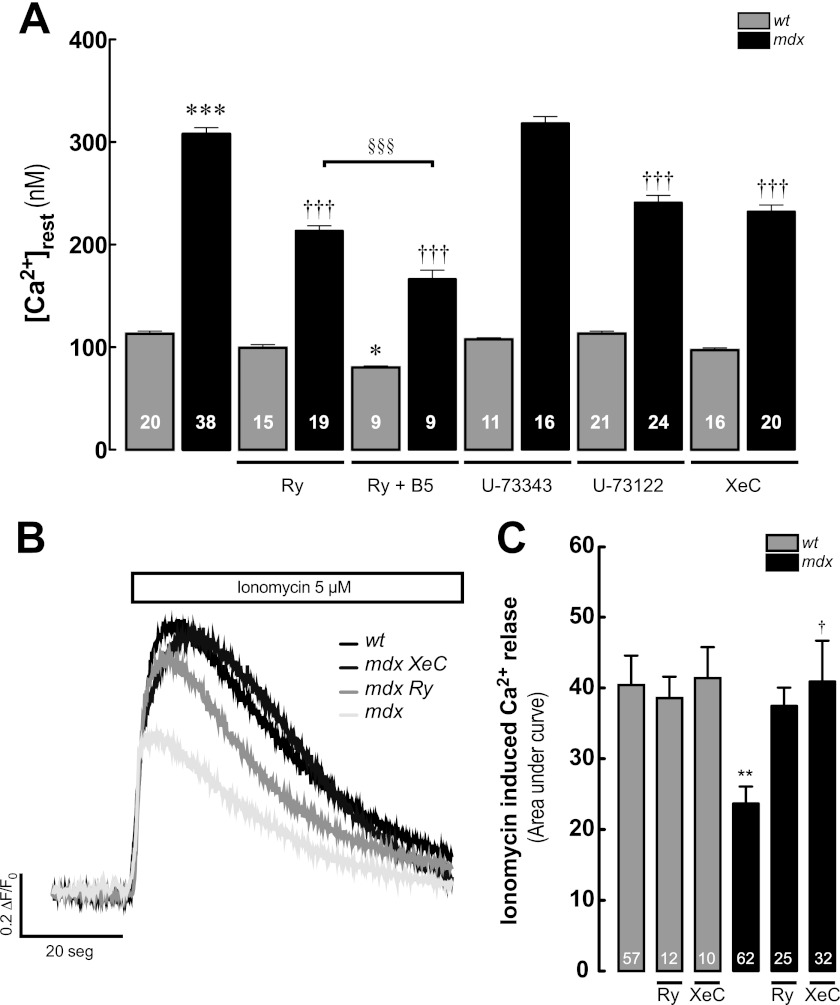
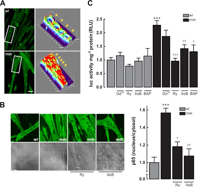
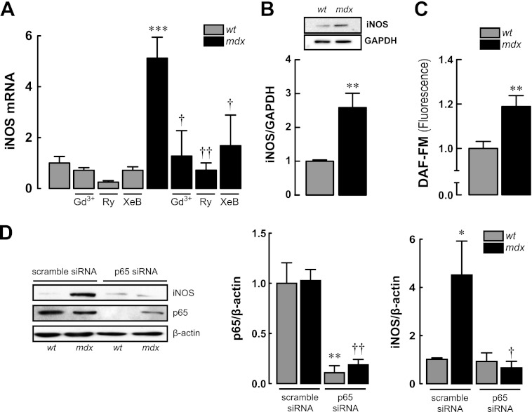
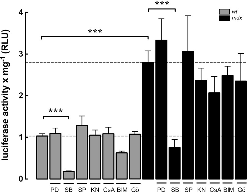
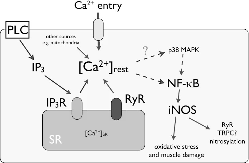
Duchenne muscular dystrophy (DMD) is a genetic disorder caused by dystrophin mutations, characterized by chronic inflammation and severe muscle wasting. Dystrophic muscles exhibit activated immune cell infiltrates, up-regulated inflammatory gene expression, and increased NF-κB activity, but the contribution of the skeletal muscle cell to this process has been unclear. The aim of this work was to study the pathways that contribute to the increased resting calcium ([Ca(2+)](rest)) observed in mdx myotubes and its possible link with up-regulation of NF-κB and pro-inflammatory gene expression in dystrophic muscle cells. [Ca(2+)](rest) was higher in mdx than in WT myotubes (308 ± 6 versus 113 ± 2 nm, p < 0.001). In mdx myotubes, both the inhibition of Ca(2+) entry (low Ca(2+) solution, Ca(2+)-free solution, and Gd(3+)) and blockade of either ryanodine receptors or inositol 1,4,5-trisphosphate receptors reduced [Ca(2+)](rest). Basal activity of NF-κB was significantly up-regulated in mdx versus WT myotubes. There was an increased transcriptional activity and p65 nuclear localization, which could be reversed when [Ca(2+)](rest) was reduced. Levels of mRNA for TNFα, IL-1β, and IL-6 were similar in WT and mdx myotubes, whereas inducible nitric-oxide synthase (iNOS) expression was increased 5-fold. Reducing [Ca(2+)](rest) using different strategies reduced iNOS gene expression presumably as a result of decreased activation of NF-κB. We propose that NF-κB, modulated by increased [Ca(2+)](rest), is constitutively active in mdx myotubes, and this mechanism can account for iNOS overexpression and the increase in reactive nitrogen species that promote damage in dystrophic skeletal muscle cells.


Figures

FIGURE 1.
FIGURE 1.
A, resting intracellular Ca2+ concentrations ([Ca2+]rest); B, resting membrane potentials (Vm) measured by double-barreled microelectrodes in WT and mdx primary myotubes. Data are expressed as means ± S.E., n = 20 for WT and n = 38 for mdx, indicated inside the bars. ***, p < 0.001 versus WT.
FIGURE 2.
FIGURE 2.
Ca2+ entry contribution to [Ca2+]rest measured in WT and mdx myotubes. A, effects of removal of extracellular Ca2+ (low Ca2+ solution and Ca2+-free solution) and Gd3+ treatment on [Ca2+]rest. B and C, measurements of resting Ca2+ entry using Mn2+ quench in WT and mdx myotubes. B, representative traces of Fura-2 fluorescence quench by Mn2+ measured under resting conditions. C, quantification of the rate of Mn2+ quench, between WT and mdx myotubes. Gd3+ (20 μm) was added during the experimental determination of Mn2+ quench as shown in figure. Data are expressed as mean ± S.E. ***, p < 0.001 versus WT basal value; †††, p < 0.001 versus mdx basal value. §§§, p < 0.001 is indicated in the figure. f.a.u./s, fluorescence arbitrary units/s.
FIGURE 3.
FIGURE 3.
RyR and IP3R participation in [Ca2+]rest in WT and mdx myotubes. A, effect of Ry (30 μm), Ry + B5 (Ry, 30 μm, and B5, 10 μm), U-73343 (5 μm), U-73122 (5 μm), and XeC (5 μm) on [Ca2+]rest (treatments for 3 h). B, representative traces of Fluo-4 fluorescence signals after the addition of 5 μm ionomycin, in the absence of extracellular Ca2+ (Ca2+-free solution) in WT, mdx, and Ry- and XeC-treated mdx myotubes. C, average area under the curve of the ionomycin-induced Ca2+ transients. Data are expressed as means ± S.E. ***, p < 0.001; **, p < 0.01; *, p < 0.05 versus WT basal value, †, p < 0.05; †††, p < 0.001 versus mdx basal value. §§§, p < 0.001 is indicated in the figure.
FIGURE 4.
FIGURE 4.
NF-κB activity in WT and mdx myotubes. A, left panel, representative z-stack immunofluorescence images obtained by confocal microscopy; right panel, three-dimensional reconstructions made with the ImageJ (National Institutes of Health) plugin Interactive 3D Surface Plot. B, effect of SR Ca2+ release inhibition in p65 subcellular distribution. C, NF-κB luciferase reporter activity in WT and mdx myotubes treated with Ca2+ inhibitors. Myotubes were incubated for 6 h and then lysed for luciferase activity determination. Data are expressed as mean ± S.E. from at least three different determinations, ***, p < 0.001 versus WT basal value, †††, p < 0.001; ††, p < 0.01 and †, p < 0.05 versus mdx basal value.
FIGURE 5.
FIGURE 5.
iNOS expression in WT and mdx myotubes. A, iNOS mRNA levels assessed by real time PCR showing effects of [Ca2+]rest reduction on iNOS mRNA expression. B, iNOS protein expression determined by Western blot. C, levels of nitric oxide (NO) was determined with DAF-FM fluorescence probe with confocal microscopy. D, effects of p65 knockdown by siRNA in the levels of p65 and iNOS proteins expression determined by Western blot. Myoblasts were transfected and then differentiated to myotubes for 48 h before the protein determination. Data are expressed as means ± S.E. from at least three different determinations. ***, p < 0.001; **, p < 0.01 and *, p < 0.05 versus WT basal value, ††, p < 0.01 and †, p < 0.05 versus mdx basal value.
FIGURE 6.
FIGURE 6.
NF-κB transcriptional activity is modulated by p38 MAPK activity in both WT and mdx myotubes. Myotubes were treated with PD-98059 (PD) (ERK1/2 inhibitor, 10 μm), SB-203580 (SB) (p38 inhibitor, 10 μm), SP-600125 (SP) (JNK inhibitor, 10 μm), KN-93 (KN) Ca2+/calmodulin-dependent kinase II (CaMKII, 10 μm), cyclosporin A (CsA) (10 μm), bisindolylmaleimide I (BIM-I) (PKC inhibitor, 2.5 μm), and Gö-6976 () ( specific inhibitor of calcium-responsive PKCs, 10 μm) for 6 h and then lysed for luciferase activity determination. Data are expressed as mean ± S.E. from at least three different determinations ***, p < 0.001.
FIGURE 7.
FIGURE 7.
Proposed model for [Ca2+]rest deregulation, NF-κB up-regulation, and iNOS expression in mdx myotubes. In addition to the Ca2+ entry through reported TRPC1 and SOCE (Gd3+ sensitive), [Ca2+]rest deregulation in mdx myotubes is a complex event that involves Ca2+ entry and SR Ca2+ leak through RyR ad IP3R. The data collected in this work suggest that increased [Ca2+]rest, increases NF-κB and iNOS expression in dystrophic myotubes. PLC, phospholipase C.

References

    1. Blake D. J., Weir A., Newey S. E., Davies K. E. (2002) Function and genetics of dystrophin and dystrophin-related proteins in muscle. Physiol. Rev. 82, 291–329 - PubMed
    1. Emery A. E. (2002) The muscular dystrophies. Lancet 359, 687–695 - PubMed
    1. Henry M. D., Campbell K. P. (1996) Dystroglycan. An extracellular matrix receptor linked to the cytoskeleton. Curr. Opin. Cell Biol. 8, 625–631 - PubMed
    1. Pasternak C., Wong S., Elson E. L. (1995) Mechanical function of dystrophin in muscle cells. J. Cell Biol. 128, 355–361 - PMC - PubMed
    1. Petrof B. J., Shrager J. B., Stedman H. H., Kelly A. M., Sweeney H. L. (1993) Dystrophin protects the sarcolemma from stresses developed during muscle contraction. Proc. Natl. Acad. Sci. U.S.A. 90, 3710–3714 - PMC - PubMed

Publication types

MeSH terms

LinkOut - more resources