Review
. 2012 Jun;21(6):751-68.
doi: 10.1002/pro.2069. Epub 2012 May 1.

The bacterial outer membrane β-barrel assembly machinery

Affiliations
Review

The bacterial outer membrane β-barrel assembly machinery

Kelly H Kim et al. Protein Sci. 2012 Jun.

Abstract

β-Barrel proteins found in the outer membrane of Gram-negative bacteria serve a variety of cellular functions. Proper folding and assembly of these proteins are essential for the viability of bacteria and can also play an important role in virulence. The β-barrel assembly machinery (BAM) complex, which is responsible for the proper assembly of β-barrels into the outer membrane of Gram-negative bacteria, has been the focus of many recent studies. This review summarizes the significant progress that has been made toward understanding the structure and function of the bacterial BAM complex.


Figures

Figure 1
Figure 1
Outer membrane proteins (OMPs) are found predominantly as β-barrels. A: Some examples of OMPs from different Gram-negative bacteria are shown. OMPs can be found either as a monomer (e.g., Pla from Yersinia pestis; PDB: 2X55), an oligomer where each subunit creates its own β-barrel (e.g., OmpLA from Escherichia coli and OprP from Pseudomonas aeruginosa; PDB: 1QD6 and 2O4V), or an oligomer where the multiple subunits come together to form one β-barrel (e.g., TolC from E. coli; PDB: 1EK9). B: OMPs share a conserved sequence of hydrophobic and aromatic residues at their C-terminus. A sequence alignment of the last 20 residues of E. coli OMPs PhoE (UniProt ID: P02932), BtuB (P06129), FadL (P10384), OmpT (P09169), OmpC (P06996), OmpF (P02931), OmpLA (P0A921), BglH (P26218), and LamB (B7UPJ7) is shown. [Color figure can be viewed in the online issue, which is available at wileyonlinelibrary.com.]
Figure 2
Figure 2
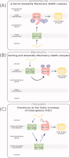
The E. coli BAM complex and homologous systems. In both Gram-negative bacteria and eukaryotes, outer membrane β-barrel proteins are first synthesized in the cytosol of the cell and then targeted to either the inner membrane (bacteria) or the proper organelle (mitochondria or chloroplasts). This figure compares the three pathways as the unfolded substrate protein (yellow curve) is directed by associated translocons (green) to the assembly complex consisting of the core BamA homologue (pink) and accessory proteins (purple), to form the final folded β-barrel (yellow cylinder). For simplicity, other proteins and chaperones involved in the pathways are not shown. A: The E. coli β-barrel assembly machinery (BAM) complex consists of membrane embedded BamA, and four accessory lipoproteins: BamB, C, D, and E. Substrate proteins cross the inner membrane via the Sec translocase and travel through the periplasmic space before being assembled by the BAM complex at the outer membrane. B: In the mitochondrial system, the substrate proteins enter via the translocase of outer mitochondrial membrane (TOM) and are assembled by the sorting and assembly machinery (SAM) complex. The BamA homologue is Sam50, which works together with cytosolic proteins Sam35 and Sam37 for insertion of OMPs into the outer mitochondrial membrane. C: In chloroplasts, the translocons at the outer and inner envelopes of chloroplasts (TOC/TIC complexes) are believed to be involved in assembly of OMPs. The BamA homologue is Toc75-V, with accessory proteins yet to be identified. It is unclear if the substrate proteins travel to the stroma before being assembled (blue arrows) or if they are directly assembled into the outer envelope membrane from the cytosol (red arrow). [Color figure can be viewed in the online issue, which is available at wileyonlinelibrary.com.]
Figure 3
Figure 3
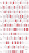
Sequence alignment of BamA. A sequence alignment is shown of E. coli BamA (UniProt ID: P0A940) with homologues from Yersinia pestis (Q8ZH58), Vibrio cholerae (Q9KPW0), Haemophilus influenzae (O32625), and Pseudomonas aeruginosa (Q9HXY4). Red boxes show absolutely conserved residues, red text shows similar residues, and blue boxes show stretches of similar residues. The secondary structure of the POTRA domains is shown above the sequence. [Color figure can be viewed in the online issue, which is available at wileyonlinelibrary.com.]
Figure 4
Figure 4
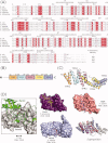
Structural features of BamA. BamA forms the core of the BAM complex and is the only component that spans the outer membrane. A: The domain structure of BamA shows an N-terminal periplasmic region that contains five POTRA domains along with a C-terminal transmembrane domain. The exact boundaries of the transmembrane domain are not known as the structure has yet to be determined, but the β-barrel is predicted to begin around Ser425. B: Ribbon diagrams of the POTRA1-4 and POTRA4-5 domains are shown. The structures for POTRA1-4 show the possibility for bent and extended conformations. C: A close up view of POTRA3 is shown, where an exposed β-strand is involved in making a crystal contact. This suggests the possibility of POTRA3 interacting with substrates or other BAM components via a process known as β-augmentation. D: The structure of FhaC from Bordetella pertussis shows the presence of only two POTRA domains, as well as a helix that can insert itself inside the β-barrel. The conserved long loop is highlighted in red. E: A domain structure comparison is shown for the BamA homologues from bacteria and eukaryotes. BamA (Escherichia coli, UniProt ID: P0A940) and Omp85 (Neisseria meningitidis, Q9K1H0) have five POTRA domains, whereas Toc75-V (in chloroplasts of Arabidopsis thaliana, Q9C5J8) has three, FhaC (Bordetella pertussis, P35077) has two, and Sam50 (in mitochondria of Saccharomyces cerevisiae, P53969) only has one. Cluster analysis of the different POTRA sequences suggests that POTRA1 of BamA, Omp85, and Toc75-V are closely related (red), whereas POTRA1 of FhaC is similar to POTRA2 of Toc75-V (cyan). The POTRA domains nearest to the membrane, for all homologues except FhaC, are closely related to each other (purple). The important VRGY motif seen in the transmembrane domain of FhaC is also conserved among the other homologues. [Color figure can be viewed in the online issue, which is available at wileyonlinelibrary.com.]
Figure 5
Figure 5
Structural features of BamB. A: A sequence alignment is shown of E. coli BamB (UniProt ID: P77774) with homologues from Salmonella typhimurium (F5ZUU9), Yersinia pestis (G0JC13), Proteus mirabilis (C2LHZ8), and Vibrio cholerae (F9ADD9). Red boxes show absolutely conserved residues, red text shows similar residues, and blue boxes show stretches of similar residues. The secondary structure is shown above the sequence. B: The domain structure of BamB shows the presence of eight domains that together form the β-propeller structure. Note that residues 46–50 form a strand that is a part of blade 8, both shown in gray. C: Ribbon diagram of BamB shows the eight-bladed β-propeller structure, with each blade numbered as in (B). The N- and C- terminus come to together to form blade 8 (gray). D: Conserved residues are shown as spheres and are found clustered on one face of the β-propeller structure. These residues are believed to be important for interaction with BamA. [Color figure can be viewed in the online issue, which is available at wileyonlinelibrary.com.]
Figure 6
Figure 6
Structural features of BamC. A: A sequence alignment is shown of E. coli BamC (UniProt ID: P0A903) with homologues from Salmonella typhi (Q83T79), Klebsiella pneumoniae (B5XVM8), Yersinia pestis (D1TUX3), and Vibrio cholerae (Q9KQ48). Red boxes show absolutely conserved residues, red text shows similar residues, and blue boxes show stretches of similar residues. The secondary structure is shown above the sequence. B: The domain structure of BamC shows the presence of three domains: an unstructured region at the N-terminus followed by two domains known as the N-terminal domain and the C-terminal domain. The C-terminal domain (pink) was solved separately and shows to have a similar helix-grip fold as the N-terminal domain (green). C: The BamC unstructured region (light blue) and N-terminal domain (green) was co-crystallized with BamD. The resulting structure shows the unstructured region to form a long loop that interacts with BamD (shown in beige). [Color figure can be viewed in the online issue, which is available at wileyonlinelibrary.com.]
Figure 7
Figure 7
Structural features of BamD. A: A sequence alignment is shown of E. coli BamD (UniProt ID: P0AC02) with homologues from Yersinia pestis (G0JE21), Candidatus Regiella insecticola (E0WTN2), Vibrio cholerae (D0I076), and Haemophilus influenzae (E4QXC3). Red boxes show absolutely conserved residues, red text shows similar residues, and blue boxes show stretches of similar residues. The secondary structure is shown above the sequence. B: The domain structure of BamD shows the presence of five TPR motifs. C: The ribbon diagram of the BamD structure is shown. Ten helices form the five TPR motifs which are numbered as in (B). D: (Left) A close up of the BamCD binding site is shown, where BamC (green) binds to a region of BamD (gray) that superimposes with the binding sites of the structural homologues. (Right) Superposition of BamD (gray), PcrH from Pseudomonas aeruginosa (purple), HOP from Homo sapiens (pink), and PEX5 from Homo sapiens (blue) show high structural similarity. These structural homologues are involved in binding to protein targeting sequences. The binding sites of these proteins are individually shown as surface models with substrate peptides as stick models. [Color figure can be viewed in the online issue, which is available at wileyonlinelibrary.com.]
Figure 8
Figure 8
Structural features of BamE. A: A sequence alignment is shown of E. coli BamE (UniProt ID: P0A937) with homologues from Salmonella typhi (Q8XF17), Yersinia pestis (G0JGU1), Haemophilus influenzae (P44057), and Vibrio cholerae (P0C6Q9). Red boxes show absolutely conserved residues, red text shows similar residues, and blue boxes show stretches of similar residues. The secondary structure is shown above the sequence. B: The ribbon diagram of a BamE monomer shows the presence of an unstructured N-terminus as well as a long flexible loop between strands β1 and β2. C: The ribbon diagram of a BamE dimer shows how the two monomers (shown in pink and green) exchange α-helices to form a domain-swapped dimer. D: The residues previously identified to be important for lipid binding are shown in yellow, and those important for BamD interaction are shown in dark pink on the surface diagrams of the BamE monomer (left) and the dimer (right). The residues shown in dark gray are important for both lipid and BamD interactions. The protein structures are shown in the same orientations as in (B) and (C). [Color figure can be viewed in the online issue, which is available at wileyonlinelibrary.com.]
Figure 9
Figure 9
Models of β-barrel assembly. Four different models of how the BAM complex may facilitate the folding and insertion of OMPs are shown. BamA is shown in pink, and the substrate protein is shown in yellow. The lipoproteins BamB/C/D/E are not shown in these models for clarity. The outer membrane is represented by the gray rectangle, with the extracellular space above and the periplasmic space below. A: In the first model, the substrate protein is first translocated across the outer membrane through the channel formed by the β-barrel domain of BamA. The substrate then inserts and folds into the outer membrane lipid bilayer from outside the cell. B: In the second model, the substrate inserts into the lipid bilayer from the periplasmic face of the outer membrane. Instead of using the channel of BamA, the insertion and folding of OMPs occur at the BamA-lipid interface. In this model, the outer wall of the β-barrel of BamA provides a scaffold for the substrate folding. C: This model is similar to the second model, but assumes that BamA forms an oligomeric structure. The coordinated events of substrate folding and membrane insertion are contained within the space formed by the BamA tetramer. The mature, folded OMP substrate is then released laterally into the lipid bilayer of the outer membrane. D: In the last model, the OMP substrate uses the N- and the C-terminal β-strands of BamA as folding templates. The hydrogen bonds between the two terminal β-strands of BamA are displaced by the incoming substrate that becomes part of the BamA structure as it folds into a β-sheet. The β-sheet of the substrate is then closed to form a β-barrel, and the substrate is released into the lipid bilayer. [Color figure can be viewed in the online issue, which is available at wileyonlinelibrary.com.]

References

    1. Silhavy TJ, Kahne D, Walker S. The bacterial cell envelope. Cold Spring Harb Perspect Biol. 2010;2(a000414):1–16. - PMC - PubMed
    1. Ricci DP, Silhavy TJ. The Bam machine: a molecular cooper. Biochim Biophys Acta. 2012;1818:1067–1084. - PMC - PubMed
    1. Fairman JW, Noinaj N, Buchanan SK. The structural biology of beta-barrel membrane proteins: a summary of recent reports. Curr Opin Struct Biol. 2011;21:523–531. - PMC - PubMed
    1. Voulhoux R, Bos MP, Geurtsen J, Mols M, Tommassen J. Role of a highly conserved bacterial protein in outer membrane protein assembly. Science. 2003;299:262–265. - PubMed
    1. Werner J, Misra R. YaeT (Omp85) affects the assembly of lipid-dependent and lipid-independent outer membrane proteins of Escherichia coli. Mol Microbiol. 2005;57:1450–1459. - PubMed

Publication types

MeSH terms

Substances

LinkOut - more resources