. 2012 Jul;53(7):1304-15.
doi: 10.1194/jlr.M025320. Epub 2012 May 1.

Evidence for the importance of OxPAPC interaction with cysteines in regulating endothelial cell function

Affiliations

Evidence for the importance of OxPAPC interaction with cysteines in regulating endothelial cell function

James R Springstead et al. J Lipid Res. 2012 Jul.

Abstract

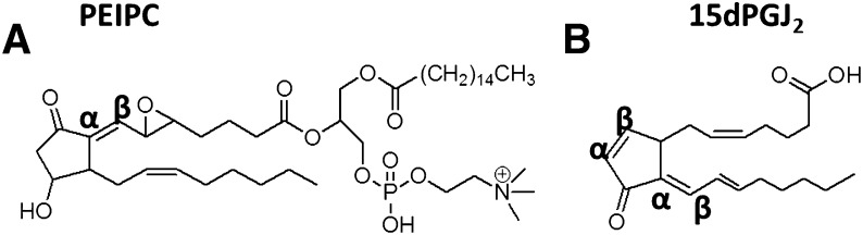
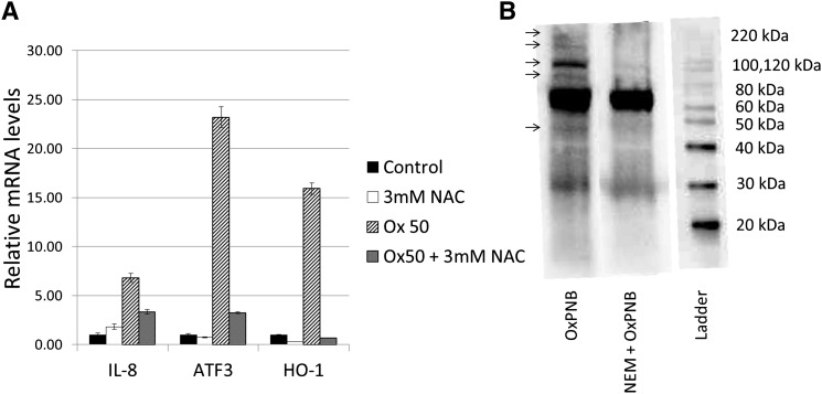
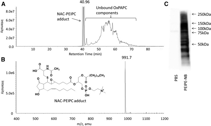
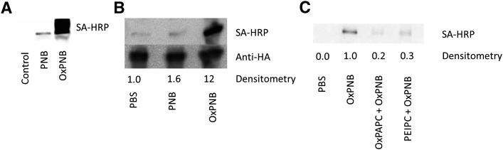
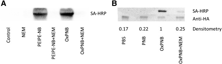
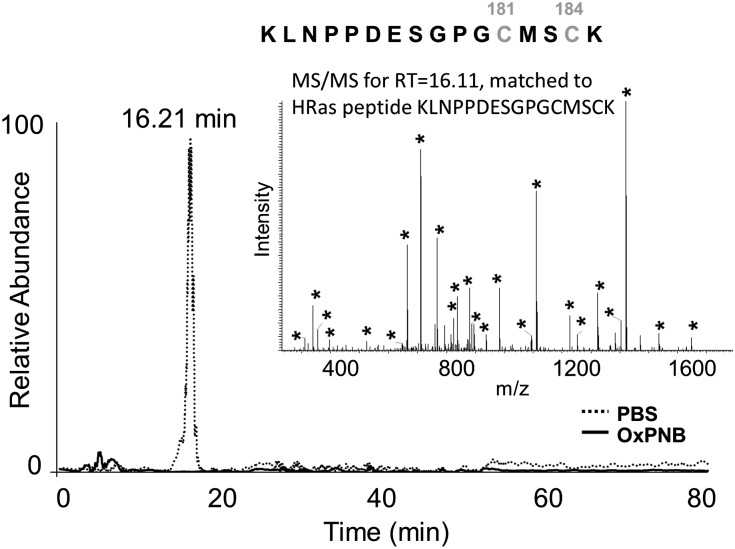
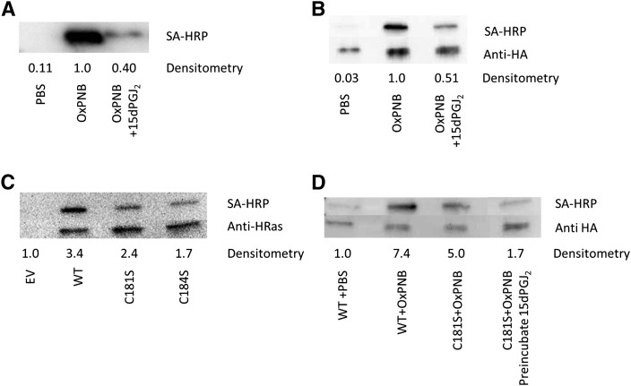
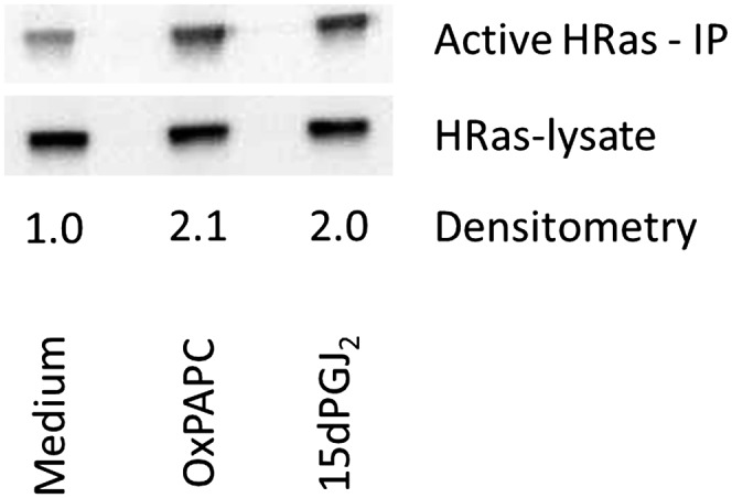
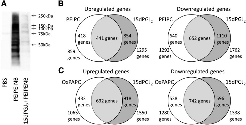
Oxidation products of 1-palmitoyl-2-arachidonoyl-sn-glycerol-3-phosphatidylcholine (PAPC), referred to as OxPAPC, and an active component, 1-palmitoyl-2-(5,6-epoxyisoprostane E₂)-sn-glycero-3-phosphatidylcholine (PEIPC), accumulate in atherosclerotic lesions and regulate over 1,000 genes in human aortic endothelial cells (HAEC). We previously demonstrated that OxPNB, a biotinylated analog of OxPAPC, covalently binds to a number of proteins in HAEC. The goal of these studies was to gain insight into the binding mechanism and determine whether binding regulates activity. In whole cells, N-acetylcysteine inhibited gene regulation by OxPAPC, and blocking cell cysteines with N-ethylmaleimide strongly inhibited the binding of OxPNB to HAEC proteins. Using MS, we demonstrate that most of the binding of OxPAPC to cysteine is mediated by PEIPC. We also show that OxPNB and PEIPE-NB, the analog of PEIPC, bound to a model protein, H-Ras, at cysteines previously shown to regulate activity in response to 15-deoxy-Δ12,14-prostaglandin J2 (15dPGJ₂). This binding was observed with recombinant protein and in cells overexpressing H-Ras. OxPAPC and PEIPC compete with OxPNB for binding to H-Ras. 15dPGJ₂ and OxPAPC increased H-Ras activity at comparable concentrations. Using microarray analysis, we demonstrate a considerable overlap of gene regulation by OxPAPC, PEIPC, and 15dPGJ₂ in HAEC, suggesting that some effects attributed to 15dPGJ₂ may also be regulated by PEIPC because both molecules accumulate in inflammatory sites. Overall, we provide evidence for the importance of OxPAPC-cysteine interactions in regulating HAEC function.


Figures

Fig. 1.
Fig. 1.
Molecular structures of (A) PEIPC and (B) 15-deoxy-Δ12,14-prostaglandin J2. The α, β carbons are shown, which are important in covalent binding.
Fig. 2.
Fig. 2.
PEIPC and OxPAPC regulate genes via interaction with cysteines. A: HAECs were incubated for 4 h in M199 or M199 with 50 μg/ml (64 μM) OxPAPC with or without pre- and cotreatment with 3 mM N-acetylcysteine (NAC). IL-8, ATF-3, and HO-1 mRNA levels were measured by qPCR and normalized to GAPDH levels. *P < 0.05 compared with the OxPAPC-treated sample, based on Student t-test. B: HAEC cells were pretreated with or without 1 mM N-ethylmaleimide (NEM) for 1 h, and then cotreated with 2.5 μg/ml (2.6 μM) OxPNB for 15 min. Western blot analysis was performed with streptavidin-HRP to detect biotinylated proteins. Molecular weight ladder and corresponding weights are shown on right. Arrows on the left identify bands that are substantially decreased in intensity with NEM treatment, indicating that NEM competes with OxPNB binding to these proteins. QPCR and binding experiments were performed at least three times, showing similar results. A representative experiment is shown.
Fig. 3.
Fig. 3.
PEIPC is the major component of OxPAPC that binds N-acetylcysteine. NAC (1 μg total, 50 μg/ml) was incubated for 4 h with 50 μg/ml (64 μM) OxPAPC and analyzed with reverse phase LC-MS/MS for PC-containing compounds by screening for compounds that yield a fragment daughter ion of 184 Da, molecular weight of the phosphatidylcholine headgroup. A: The chromatogram represents the total ion current of species eluting from the column. The sharp peak at 40.96 min represents the NAC-PEIPC adduct eluting from the column. The peaks that elute at later retention times are unbound residual OxPAPC species. B: The MS spectra of the peak eluting at 40.96, with a molecular weight of 991.7 Da, representing the NAC-PEIPC adduct (structure shown in inset); C: HAEC cells were lysed and either run directly or treated with 12 μM PEIPE-NB. Western blot analysis was performed with streptavidin-HRP to detect biotinylated proteins.
Fig. 4.
Fig. 4.
Oxidized PAPE-N-biotin binds to recombinant H-Ras and H-Ras in cells, whereas unoxidized PAPE-N-biotin does not bind. A: Human recombinant H-Ras (hr-H-Ras) was incubated with no lipid, 50 μg/ml (52 μM) unoxidized PAPE-N-biotin (PNB), or 50 μg/ml (52 μM) oxidized PAPE-N-biotin (OxPNB) for 30 min. Western blot analysis was performed with SA-HRP to detect biotinylated (lipid-modified) H-Ras. B: HEK293 cells were transfected with HA-tagged wild-type H-Ras. Cells were treated with PBS or 50 μg/ml (52 μM) of unoxidized or oxidized PAPE-N-biotin for 30 min. Lysates were immunoprecipitated with anti-HA beads, and samples were analyzed by Western blot for HA and streptavidin (lipid-modified H-Ras). Densitometry shows the ratio of lipid-modified H-Ras (SA-HRP) to total H-Ras (anti-HA); These experiments were performed at least three times, showing similar results. C: 100 ng hr-H-Ras was incubated with no lipid, 100 μM OxPAPC, or 10 μM PEIPC for 60 min, followed by incubation with no lipid or 10 μM OxPNB for an additional 15 min. Western blot analysis was performed as in panel A. Densitometry shows the ratio of lipid-modified H-Ras normalized to H-Ras treated only with OxPNB.
Fig. 5.
Fig. 5.
N-ethylmaleimide competes for binding of OxPAPE-N-biotin and PEIPE-N-biotin to H-Ras. A: 1 μg hr-H-Ras was incubated with no lipid, 0.2 mM NEM, 0.5 μg PEIPE-NB, 0.5 μg PEIPE-NB and 0.2 mM NEM, 0.5 μg OxPNB, or 0.5 μg OxPNB and 0.2 μg NEM for 30 min, corresponding to 10:1 NEM or lipid:H-Ras molar ratios. Western blot analysis was performed as in Fig. 3A. B: HEK293 cells were transfected with HA-tagged wild-type H-Ras and treated with PBS or 1 mM NEM for 60 min, then with 10 μg/ml (10.4 μM) OxPNB for 15 min. Lysates were prepared for Western blotting as in Fig. 3B. Densitometry shows the ratio of lipid-modified H-Ras (SA-HRP) to total H-Ras (anti-HA). Competition experiments were performed at least three times, showing similar results.
Fig. 6.
Fig. 6.
LC/MS shows that OxPAPE-N-biotin binding to H-Ras involves cysteines 181 and 184. Hr-H-Ras was incubated with or without OxPNB at a molar ratio of 1:10 (hr-H-Ras:OxPNB) at 37°C for 30 min, digested with trypsin, and analyzed with ESI-LC/MS/MS, followed by identification of H-Ras fragments. One peptide, eluting at 16.21 min, was found only in untreated H-Ras and not in OxPNB-treated H-Ras. The inset shows the MS/MS spectra of the peptide eluting at 16.2 min with the sequence KLNPPDESGPGCMSCK. Asterisks indicate peaks used for matching the MS/MS peaks to the peptide in the database. The observed m/z for this fragment is 888.87, representing a doubly charged ion of molecular weight 1775. This data shows that the binding of OxPNB to H-Ras involves C181 and/or C184.
Fig. 7.
Fig. 7.
Mutant H-Ras and 15dPGJ2 provide evidence that binding of Ox-PAPE-N-Biotin to H-Ras involves Cysteines 181 and 184. A: Ability of 15dPGJ2 to compete with OxPNB for binding to recombinant H-Ras. Human recombinant H-Ras was preincubated with either PBS or 50 μg/ml (158 μM) 15dPGJ2 for 30 min, and then coincubated with 50 μg/ml (52 μM) OxPNB for 30 min. Western blotting was performed as in Fig. 3A. B: Ability of 15dPGJ2 to compete with OxPNB for binding to H-Ras in cells. HEK293 cells were transfected with HA-tagged wild-type H-Ras and treated with PBS or 50 μg/ml (158 μM) 15dPGJ2 for 30 min, and then 50 μg/ml (52 μM) OxPNB for 30 min. Lysates were prepared for Western blotting as in Fig. 3B. Densitometry shows the ratio of lipid-modified H-Ras (SA-HRP) to total H-Ras (anti-HA). C: HEK293 cells were transfected with empty vector, HA-tagged wild-type H-Ras, C181S-mutant H-Ras, or C184S-mutant H-Ras. Cells were treated with 50 μg/ml (52 μM) OxPNB for 30 min. Lysates were immunoprecipitated with anti-HA resin, and Western blotting was carried out as in Fig. 3A. D: HEK293 cells were transfected with empty vector, HA-tagged wild-type H-Ras, or HA-tagged C181S-mutant H-Ras. Cells were treated with 10 μg/ml (10.4 μM) OxPNB for 30 min. The last lane was pretreated with 50 μg/ml (158 μM) 15dPGJ2 for 15 min, and then cotreated with 10 μg/ml (10.4 μM) OxPNB for 30 min. Cells were immunoprecipitated, and Western blotting was carried out as in Fig. 3A. Mutant and competition H-Ras experiments were performed at least three times, showing similar results.
Fig. 8.
Fig. 8.
H-Ras in HAECs is activated by both OxPAPC and 15dPGJ2. One hundred millimeter dishes of HAECs were incubated in M199 or M199 with 64 μM OxPAPC or 15dPGJ2 for 60 min. Activated H-Ras was isolated as per Ras pulldown kit manufacturer instructions (Pierce Biotechnologies). Densitometry shows the ratio of active H-Ras (after kit immunoprecipitation) to total H-Ras (before immunoprecipitation); Densitometry was performed normalized to lysate and to M199 signals. Activation data were replicated three times, showing consistent activation by OxPAPC and 15dPGJ2 at 60 min.
Fig. 9.
Fig. 9.
Evidence for an overlap in binding and regulation of gene expression in HAEC by PEIPC, OxPAPC, and 15dPGJ2. A: HAEC cells were lysed, and lysates were treated with 12 μM PEIPE-NB or cotreated with 120 μM 15dPGJ2 and 12 μM PEIPE-NB. Western blotting was then performed. This experiment was performed at least three times with similar results. B: Duplicate wells were treated with media or media containing 50 μg/ml (64 μM) OxPAPC or equimolar amounts of either PEIPC or 15dPGJ2 for 4 h, and then RNA was extracted and analyzed with microarray on an Illumina chip. Data were filtered for P ≤ 0.001 and fold change of 1.25 during data analysis with Genome Studio software; Data are represented in Venn diagrams of PEIPC versus 15dPGJ2 downregulated and upregulated genes and OxPAPC versus 15dPGJ2 downregulated and upregulated genes.

References

    1. Berliner J. A., Navab M., Fogelman A. M., Frank J. S., Demer L. L., Edwards P. A., Watson A. D., Lusis A. J. 1995. Atherosclerosis: basic mechanisms. oxidation, inflammation, and genetics. Circulation. 91: 2488–2496 - PubMed
    1. Berliner J. A., Leitinger N., Tsimikas S. 2009. The role of oxidized phospholipids in atherosclerosis. J. Lipid Res. 50(Suppl): S207–S212 - PMC - PubMed
    1. Romanoski C. E., Che N., Yin F., Mai N., Pouldar D., Civelek M., Pan C., Lee S., Vakili L., Yang W. P., et al. 2011. Network for activation of human endothelial cells by oxidized phospholipids: a critical role of heme oxygenase 1. Circ. Res. 109: e27–e41 - PMC - PubMed
    1. Gugiu B. G., Mouillesseaux K., Duong V., Herzog T., Hekimian A., Koroniak L., Vondriska T. M., Watson A. D. 2008. Protein targets of oxidized phospholipids in endothelial cells. J. Lipid Res. 49: 510–520 - PubMed
    1. Gao D., Ashraf M. Z., Kar N. S., Lin D., Sayre L. M., Podrez E. A. 2010. Structural basis for the recognition of oxidized phospholipids in oxidized low density lipoproteins by class B scavenger receptors CD36 and SR-BI. J. Biol. Chem. 285: 4447–4454 - PMC - PubMed

Publication types

MeSH terms

Substances