. 2012 May 15;109(20):7899-904.
doi: 10.1073/pnas.1201997109. Epub 2012 May 1.

Structure and stoichiometry of an accessory subunit TRIP8b interaction with hyperpolarization-activated cyclic nucleotide-gated channels

Affiliations

Structure and stoichiometry of an accessory subunit TRIP8b interaction with hyperpolarization-activated cyclic nucleotide-gated channels

John R Bankston et al. Proc Natl Acad Sci U S A. .

Abstract

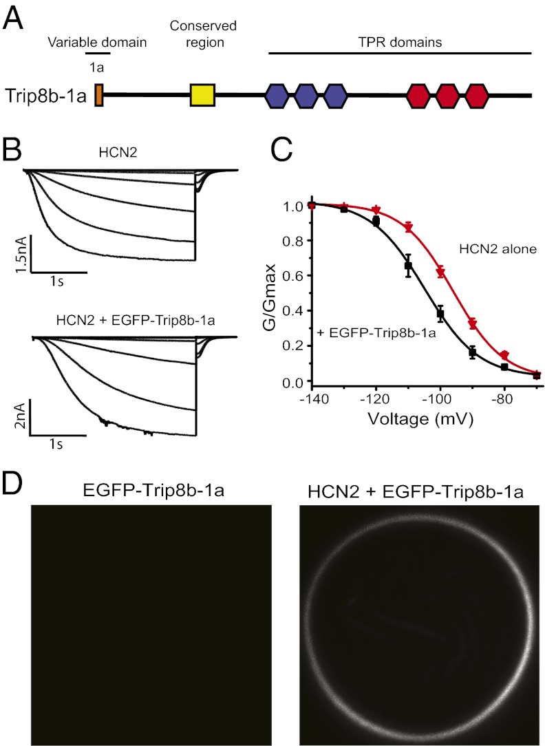
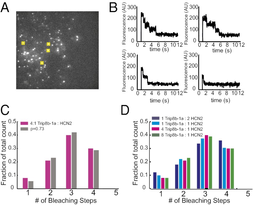
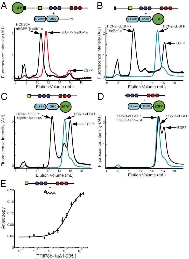
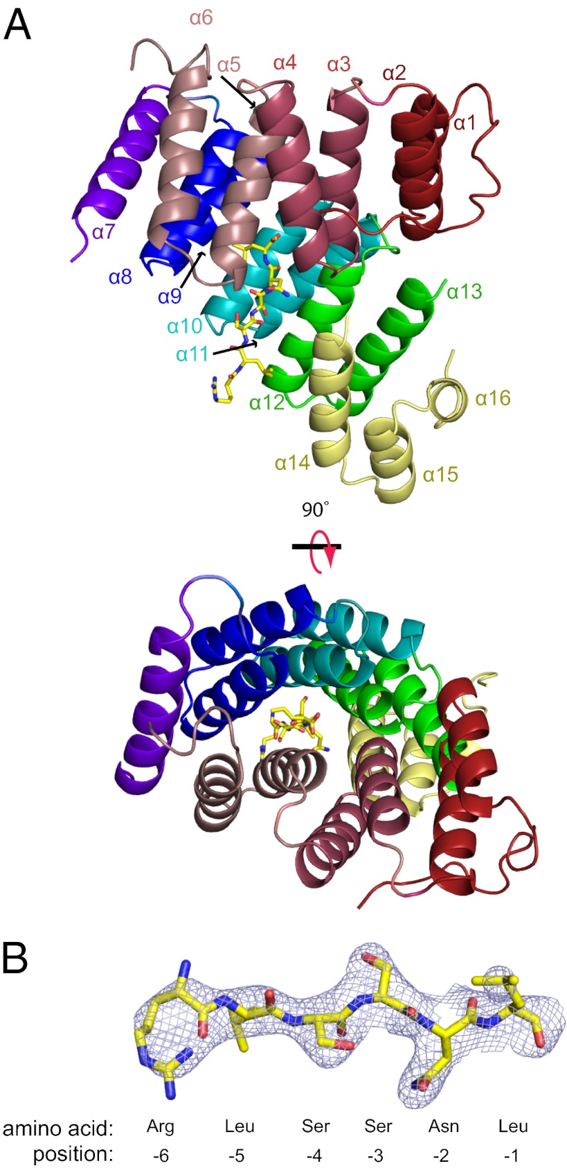
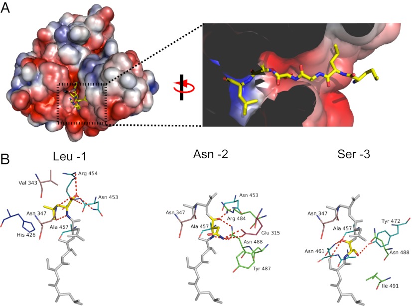
Ion channels operate in intact tissues as part of large macromolecular complexes that can include cytoskeletal proteins, scaffolding proteins, signaling molecules, and a litany of other molecules. The proteins that make up these complexes can influence the trafficking, localization, and biophysical properties of the channel. TRIP8b (tetratricopetide repeat-containing Rab8b-interacting protein) is a recently discovered accessory subunit of hyperpolarization-activated cyclic nucleotide-gated (HCN) channels that contributes to the substantial dendritic localization of HCN channels in many types of neurons. TRIP8b interacts with the carboxyl-terminal region of HCN channels and regulates their cell-surface expression level and cyclic nucleotide dependence. Here we examine the molecular determinants of TRIP8b binding to HCN2 channels. Using a single-molecule fluorescence bleaching method, we found that TRIP8b and HCN2 form an obligate 4:4 complex in intact channels. Fluorescence-detection size-exclusion chromatography and fluorescence anisotropy allowed us to confirm that two different domains in the carboxyl-terminal portion of TRIP8b--the tetratricopepide repeat region and the TRIP8b conserved region--interact with two different regions of the HCN carboxyl-terminal region: the carboxyl-terminal three amino acids (SNL) and the cyclic nucleotide-binding domain, respectively. And finally, using X-ray crystallography, we determined the atomic structure of the tetratricopepide region of TRIP8b in complex with a peptide of the carboxy-terminus of HCN2. Together, these experiments begin to uncover the mechanism for TRIP8b binding and regulation of HCN channels.


Conflict of interest statement

The authors declare no conflict of interest.

Figures

Fig. 1.
Fig. 1.
EGFP-tagged TRIP8b-1a bound to HCN2 channels regulates their voltage dependence. (A) Cartoon showing the isoform TRIP8b-1a used in this study. (B) Current families from cell-attached patches on oocytes that expressed HCN2 alone and HCN2 with EGFP-TRIP8b-1a. (C) Conductance-voltage relationship for HCN2 channels and HCN2 channels with EGFP-TRIP8b-1a measured in the cell-attached patch configuration. (D) Confocal microscopy images of oocytes expressing EGFP-TRIP8b-1a alone and EGFP-TRIP8b-1a coexpressed with HCN2 channels.
Fig. 2.
Fig. 2.
Single-molecule bleaching revealed a 4:4 TRIP8b/HCN2 stoichiometry. (A) TIRF microscopy was used to image the membranes of oocytes. The image shows an average of five frames of a representative movie of EGFP-TRIP8b-1a coexpressed with HCN2. Yellow boxes represent a sample of selected spots for analysis. (B) Examples of bleaching steps for EGFP-TRIP8b-1a associated with HCN2 channels. Each graph shows a spot that bleaches in a different number of steps from 1 to 4. (C) Distribution of bleaching steps for an RNA injection ratio of 4:1 EGFP-TRIP8b-1a:HCN2. Magenta bars show the fraction of spots that bleached in a given number of steps. Data were obtained from 508 spots. The gray bars show the fit of a binomial distribution with n = 4 and P = 0.73. (D) Distribution of bleaching steps from various injection ratios of EGFP-TRIP8b-1a and HCN2 RNA. These data were obtained from 242, 289, and 277 spots for the 8:1, 2:1, and 1:2 ratios, respectively.
Fig. 3.
Fig. 3.
TRIP8b-1a forms a bipartite interaction with HCN2. Each panel contains a cartoon showing the protein constructs used and the location of the fluorescent tag. (A) FSEC results of the crude lysates containing TRIP8b-1a (red) and TRIP8b-1a coexpressed with HCN2C (black). (B) FSEC results of the crude lysates containing HCN2I (cyan) and HCN2I coexpressed with TRIP8b-1a (black). (C) FSEC results of crude lysates containing HCN2I (cyan) and HCN2I coexpressed with TRIP8b-1aΔ1–205 (black). (D) FSEC results from crude lysates containing HCN2I (cyan) and HCN2 coexpressed with TRIP8b-1aΔ1–254 (black). (E) Fluorescence anisotropy of the TAMRA-SNL peptide plotted versus the total concentration of TRIP8b-1aΔ1–205. The data were fit with a Kd of 155 ± 31 nM (equation in Materials and Methods).
Fig. 4.
Fig. 4.
Crystal structure of TRIP8b-1aΔ1–205 in complex with a peptide of the last seven amino acids of HCN2. (A) Ribbon representation of TRIP8b-1aΔ1–205 shown from two different perspectives with the SNL peptide shown in stick representation. Each TPR is colored differently (TRP1, red; TPR2, dark pink; TRP3, light pink; TPR4, blue; TPR5, cyan; TPR6, green; hinge region helix, purple; three helix bundle, yellow). (B) Fo-Fc electron density “omit” map contoured at 1σ for the SNL peptide. In addition, the numbering system for the peptide is shown below.
Fig. 5.
Fig. 5.
Binding site for the SNL peptide on TRIP8b. (A) Molecular surface representation of TRIP8b-1aΔ1–205 colored according to the calculated electrostatic potential (−3 to 3 kTe). Close-up of the binding pocket from A with the SNL peptide in stick representation. The surface display was clipped to show the pocket where HCN2 binds. In addition, two residues (Asn-347 and Cys-355) were removed from the figure for clarity. (B) Interactions with TRIP8b broken down by residue on the SNL peptide. The colors of the carbons on TRIP8b match the color scheme of the ribbon representation in Fig. 4A. Polar contacts are shown as dashed red lines.

References

    1. Buraei Z, Yang J. The ß subunit of voltage-gated Ca2+ channels. Physiol Rev. 2010;90:1461–1506. - PMC - PubMed
    1. Pongs O, Schwarz JR. Ancillary subunits associated with voltage-dependent K+ channels. Physiol Rev. 2010;90:755–796. - PubMed
    1. Trudeau MC, Zagotta WN. Calcium/calmodulin modulation of olfactory and rod cyclic nucleotide-gated ion channels. J Biol Chem. 2003;278:18705–18708. - PubMed
    1. Craven KB, Zagotta WN. CNG and HCN channels: Two peas, one pod. Annu Rev Physiol. 2006;68:375–401. - PubMed
    1. Robinson RB, Siegelbaum SA. Hyperpolarization-activated cation currents: From molecules to physiological function. Annu Rev Physiol. 2003;65:453–480. - PubMed

Publication types

MeSH terms

Substances

Associated data