Review
. 2012 May 1;4(5):a008318.
doi: 10.1101/cshperspect.a008318.

Signaling networks regulating development of the lower respiratory tract

Affiliations
Review

Signaling networks regulating development of the lower respiratory tract

David M Ornitz et al. Cold Spring Harb Perspect Biol. .

Abstract

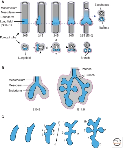
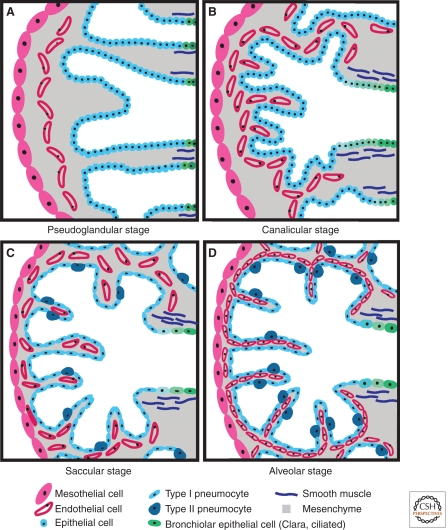
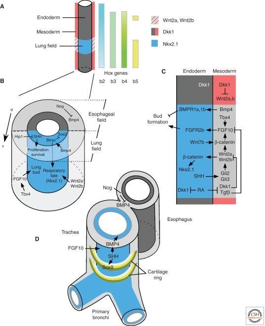
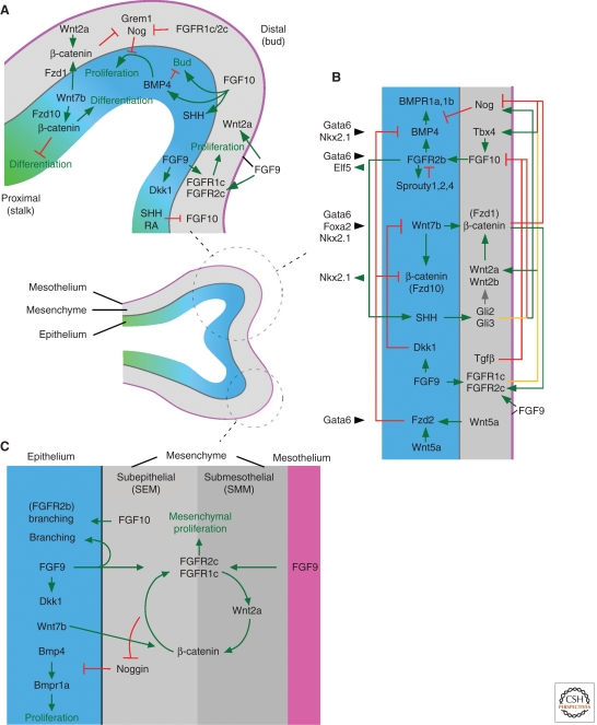
The lungs serve the primary function of air-blood gas exchange in all mammals and in terrestrial vertebrates. Efficient gas exchange requires a large surface area that provides intimate contact between the atmosphere and the circulatory system. To achieve this, the lung contains a branched conducting system (the bronchial tree) and specialized air-blood gas exchange units (the alveoli). The conducting system brings air from the external environment to the alveoli and functions to protect the lung from debris that could obstruct airways, from entry of pathogens, and from excessive loss of fluids. The distal lung enables efficient exchange of gas between the alveoli and the conducting system and between the alveoli and the circulatory system. In this article, we highlight developmental and physiological mechanisms that specify, pattern, and regulate morphogenesis of this complex and essential organ. Recent advances have begun to define molecular mechanisms that control many of the important processes required for lung organogenesis; however, many questions remain. A deeper understanding of these molecular mechanisms will aid in the diagnosis and treatment of congenital lung disease and in the development of strategies to enhance the reparative response of the lung to injury and eventually permit regeneration of functional lung tissue.


Figures

Figure 1.
Figure 1.
Morphogenesis of the bronchial tree. (A) The embryonic stage of lung development includes specification of the primary lung field and formation of lung buds (primary bronchi) and trachea. The foregut tube is composed of endoderm, mesoderm, and the mesothelium (purple line). The mesothelium is an epithelial-like cell layer of mesodermal-derived cells that lines the surface of visceral organs within the coelomic cavity. Lung buds appear on the ventral (V) foregut at the 24 somite (24S) stage (E9.75 in the mouse). Before the appearance of lung buds, Nkx2.1 is expressed in ventral foregut endoderm (blue), marking the primary lung field. The tracheoesophageal groove (arrowhead) initiates septation of the anterior foregut and esophagus. The mesothelium associated with the lung buds (pink) will eventually give rise to the visceral pleura. Corresponding somite stages and embryonic ages are indicated. Arrowhead indicates the tracheoesophageal groove. Dashed line indicates the plane of cross section shown below and on the right. v, ventral; d, dorsal. (Adapted from Spooner and Wessells 1970). (B) Branching morphogenesis continues throughout the pseudoglandular stage (E10.5-E16.5 in the mouse) and gives rise to conducting airways and alveolar sacs. (C) Primary lung lobes are formed by domain branching mechanisms and secondary subdivisions are formed by planar bifarcations. Domain branching regulates the temporal formation of buds along the proximal (p)–distal (d) axis (1–5) and their position on a circumferential axis (c). Domain branching is followed by orthogonal and planar bifurcations (1b) leading to iterative expansion of the endodermal structure of the lung.
Figure 2.
Figure 2.
Stages of lung development. (A) The pseudoglandular stage (mouse E10.5 to 16.5; human E52 to week 17) begins with formation of secondary bronchi and includes formation of conducting airways, a primitive capillary plexus, and differentiation of mesenchyme to form smooth muscle. Histologically, this stage is characterized by glandularlike structures separated by abundant mesenchyme. (B) The canalicular stage (mouse E16.5 to 17.5; human wk 17 to 28) is characterized by thinning of the distal epithelium to form primitive pulmonary acini (terminal sacs), early differentiation of type I and type II pneumocytes (alveolar epithelial cells), and continued angiogenesis and juxtaposition of capillaries with the respiratory epithelium to increase gas exchange surface area. Mesenchyme in the canalicular stage is still relatively abundant. (C) In the saccular stage (mouse E17.5 to postnatal (P) day 5; human wk 28 to 36), the terminal epithelial sacs continue to separate and become enveloped by capillaries. Type II pneumocytes begin to produce surfactant and continue to differentiate into type I pneumocytes. The submesothelial and subepithelial mesenchyme becomes thinner and contains more differentiated cells, including bronchiolar and vascular smooth muscle. (D) During the alveolar stage (mouse P5 to 30; human wk 36 to 40), the terminal epithelial sacs continue to form primary and secondary septa, creating mature alveoli.
Figure 3.
Figure 3.
Signaling pathways regulating respiratory derivatives of the foregut. (A) Mesenchymal and endodermal molecules that pattern the foregut and lung field. (B) Signaling molecules and key transcription factors that regulate lung bud formation. (C) Signaling networks within and between endodermal and mesodermal components of the foregut that regulate lung bud formation. (D) Signaling molecules that regulate morphogenesis of the trachea and esophagus.
Figure 4.
Figure 4.
Signaling pathways regulating lung development at the early (E12.5) pseudoglandular stage. (Center) Diagram showing a branching epithelial tube (distal, blue and proximal, green) surrounded by mesenchyme (gray) and mesothelium (pink). (A) Signaling molecules that regulate growth, budding, and differentiation of the airway epithelium and surrounding mesenchyme. (B) Signaling network model of the distal lung bud. (C) Feed-forward signaling network within subepithelial and submesothelial mesenchyme.
Figure 5.
Figure 5.
Molecular markers expressed in pseudoglandular stage lung mesenchyme. (A) Wnt2a expression in submesothelial mesenchyme. (B) Noggin-LacZ expression in subepithelial mesenchyme. (C) Endothelial lineage marked by Flk1-Cre activation of the R26R reporter gene. (D) Smooth muscle actin (Sma) expression in peribronchiolar and perivascular tissue. (E) Mesodermal and mesothelial lineage marked by Dermo1-Cre activation of the R26R reporter gene. (F) Epithelial lineages marked by Shh-Cre activation of the R26R reporter gene. e, epithelium; m, mesenchyme; meso, mesothelium; v, blood vessel.

References

    1. Abadie V, Champagnat J, Fortin G 2000. Branchiomotor activities in mouse embryo. Neuroreport 11: 141–145 - PubMed
    1. Abler LL, Mansour SL, Sun X 2009. Conditional gene inactivation reveals roles for Fgf10 and Fgfr2 in establishing a normal pattern of epithelial branching in the mouse lung. Dev Dyn 238: 1999–2013 - PMC - PubMed
    1. Ahlbrecht K, Schmitz J, Seay U, Schwarz C, Mittnacht-Kraus R, Gaumann A, Haberberger RV, Herold S, Breier G, Grimminger F, et al. 2008. Spatiotemporal expression of flk-1 in pulmonary epithelial cells during lung development. Am J Respir Cell Mol Biol 39: 163–170 - PubMed
    1. Aubin J, Lemieux M, Tremblay M, Berard J, Jeannotte L 1997. Early postnatal lethality in Hoxa-5 mutant mice is attributable to respiratory tract defects. Dev Biol 192: 432–445 - PubMed
    1. Baguma-Nibasheka M, Angka HE, Inanlou MR, Kablar B 2007. Microarray analysis of Myf5−/−:MyoD−/− hypoplastic mouse lungs reveals a profile of genes involved in pneumocyte differentiation. Histol Histopathol 22: 483–495 - PubMed

Publication types

MeSH terms

LinkOut - more resources