Review
. 2012 May 1;4(5):a011536.
doi: 10.1101/cshperspect.a011536.

The structure and function of the eukaryotic ribosome

Affiliations
Review

The structure and function of the eukaryotic ribosome

Daniel N Wilson et al. Cold Spring Harb Perspect Biol. .

Abstract

Structures of the bacterial ribosome have provided a framework for understanding universal mechanisms of protein synthesis. However, the eukaryotic ribosome is much larger than it is in bacteria, and its activity is fundamentally different in many key ways. Recent cryo-electron microscopy reconstructions and X-ray crystal structures of eukaryotic ribosomes and ribosomal subunits now provide an unprecedented opportunity to explore mechanisms of eukaryotic translation and its regulation in atomic detail. This review describes the X-ray crystal structures of the Tetrahymena thermophila 40S and 60S subunits and the Saccharomyces cerevisiae 80S ribosome, as well as cryo-electron microscopy reconstructions of translating yeast and plant 80S ribosomes. Mechanistic questions about translation in eukaryotes that will require additional structural insights to be resolved are also presented.


Figures
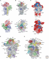
Figure 1.
Figure 1.
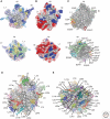
The bacterial and eukaryotic small ribosomal subunit. (A,B) Interface (upper) and solvent (lower) views of the bacterial 30S subunit (Jenner et al. 2010a). (A) 16S rRNA domains and associated r-proteins colored distinctly: b, body (blue); h, head (red); pt, platform (green); and h44, helix 44 (yellow). (B) 16S rRNA colored gray and r-proteins colored distinctly and labeled. (CE) Interface and solvent views of the eukaryotic 40S subunit (Rabl et al. 2011), with (C) eukaryotic-specific r-proteins (red) and rRNA (pink) shown relative to conserved rRNA (gray) and r-proteins (blue), and with (D,E) 18S rRNA colored gray and r-proteins colored distinctly and labeled.
Figure 2.
Figure 2.
The bacterial and eukaryotic large ribosomal subunit. (A) Interface (upper) and solvent (lower) views of the bacterial 50S subunit (Jenner et al. 2010b), with 23S rRNA domains and bacterial-specific (light blue) and conserved (blue) r-proteins colored distinctly: cp, central protuberance; L1, L1 stalk; and St, L7/L12 stalk (or P-stalk in archeaa/eukaryotes). (BE) Interface and solvent views of the eukaryotic 60S subunit (Klinge et al. 2011), with (B) eukaryotic-specific r-proteins (red) and rRNA (pink) shown relative to conserved rRNA (gray) and r-proteins (blue), (C) eukaryotic-specific expansion segments (ES) colored distinctly, and (D,E) 28S rRNA colored gray and r-proteins colored distinctly and labeled.
Figure 3.
Figure 3.
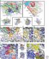
Structural and functional aspects of the eukaryotic ribosome. Interweaving of rRNA and r-proteins on the (A) LSU near ES7L and ES39L (Klinge et al. 2011), and (B) SSU near ES3 and ES6 (Rabl et al. 2011). Extension of r-proteins at the tRNA-binding sites on the (C) SSU (Armache et al. 2010b; Rabl et al. 2011), LSU of the (D) bacterial (Jenner et al. 2010b), and (E) eukaryotic (Armache et al. 2010b) peptidyltransferase centers. R-proteins located at the mRNA (F) exit, and (G) entry sites (Klinge et al. 2011).
Figure 4.
Figure 4.
Positioning of eIF1 near the SSU P site. (A) Steric clash between eIF1 and P-site tRNA in the canonical P/P configuration. Structure of the 40S subunit–eIF1 complex superimposed with the unrotated state of the ribosome in Dunkle et al. (2011). (B) Binding of eIF1 is more compatible with tRNA in the P/E configuration. Structure of the 40S subunit–eIF1 complex superimposed with the rotated state of the ribosome in Dunkle et al. (2011). Nucleotides in 18S rRNA that would contribute to contacts with the LSU in bridge B2a are colored red.
Figure 5.
Figure 5.
Intersubunit rotation required for translation. (A) Key conformational rearrangements in the ribosome. Rotation of the SSU body, head domain, and opening of the mRNA- and tRNA-binding groove during mRNA and tRNA translocation (asterisk) are indicated by arrows. Closing of the SSU body toward the LSU during mRNA decoding is also indicated by an arrow. Dynamic regions of the LSU (L1 arm, P proteins, and GTPase center) are labeled. (B) Bridges eB12 and eB13 in the yeast ribosome at the periphery of the subunits. LSU proteins contributing to the bridges are marked. The view is indicated to the left. (C) Bridge eB14 in the yeast ribosome, near the pivot point of intersubunit rotation. LSU protein L41e and 18S rRNA helices in the SSU contributing to the bridge (gold) are indicated.

References

    1. Alkalaeva EZ, Pisarev AV, Frolova LY, Kisselev LL, Pestova TV 2006. In vitro reconstitution of eukaryotic translation reveals cooperativity between release factors eRF1 and eRF3. Cell 125: 1125–1136 - PubMed
    1. Allen GS, Zavialov A, Gursky R, Ehrenberg M, Frank J 2005. The cryo-EM structure of a translation initiation complex from Escherichia coli. Cell 121: 703–712 - PubMed
    1. Andersen GR, Pedersen L, Valente L, Chatterjee I, Kinzy TG, Kjeldgaard M, Nyborg J 2000. Structural basis for nucleotide exchange and competition with tRNA in the yeast elongation factor complex eEF1A:eEF1Bα. Mol Cell 6: 1261–1266 - PubMed
    1. Andersen CB, Becker T, Blau M, Anand M, Halic M, Balar B, Mielke T, Boesen T, Pedersen JS, Spahn CM, et al. 2006. Structure of eEF3 and the mechanism of transfer RNA release from the E-site. Nature 443: 663–668 - PubMed
    1. Armache JP, Jarasch A, Anger AM, Villa E, Becker T, Bhushan S, Jossinet F, Habeck M, Dindar G, Franckenberg S, et al. 2010a. Cryo-EM structure and rRNA model of a translating eukaryotic 80S ribosome at 5.5-Å resolution. Proc Natl Acad Sci 107: 19748–19753 - PMC - PubMed

Publication types

MeSH terms

LinkOut - more resources