

Transplantation of amniotic membrane to the subretinal space in pigs
- PMID: 22550516
- PMCID: PMC3328183
- DOI: 10.1155/2012/716968
Transplantation of amniotic membrane to the subretinal space in pigs
Abstract
Purpose: To investigate the effect of transplanted amniotic membrane (AM) on subretinal wound healing.
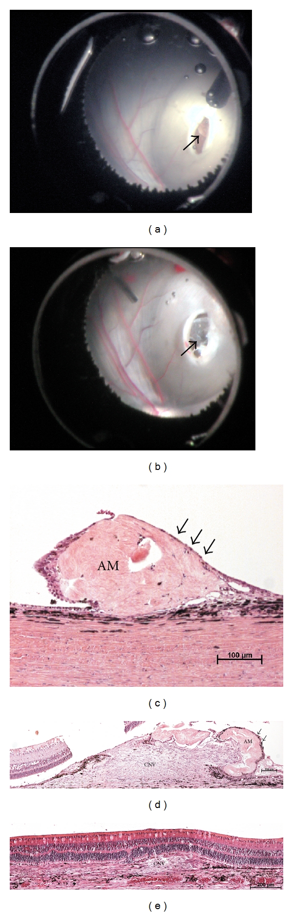
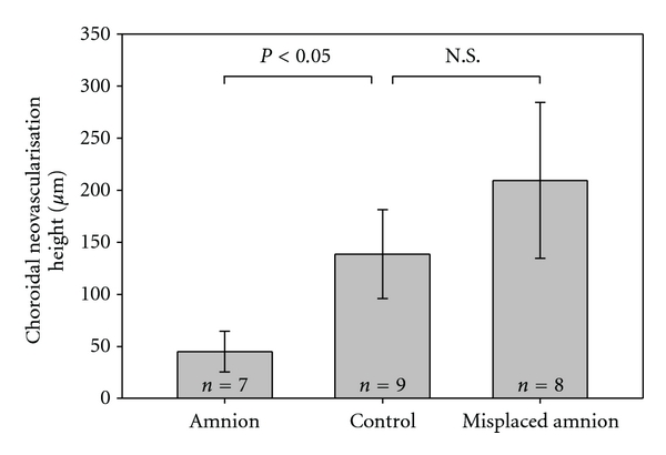
Methods: Nine Danish Landrace pigs had surgical removal of retinal pigment epithelium (RPE) and mechanical damage of Bruch's membrane (BM) and served as a control group. 15 pigs additionally had AM transplanted to the subretinal space.
Results: AM significantly reduces choroidal neovascularisation when complete coverage of the induced defect is obtained (7 pigs) (P < 0.05). In cases where AM did not cover the rupture in BM choroidal tissue covered the transplanted membrane (8 pigs). AM is well tolerated in the subretinal space, causes only limited inflammation, and is covered with a monolayer of pigmented cells when in contact with the host RPE.
Conclusions: AM modifies choroidal neovascularisation after BM damage and may serve as a basement membrane substitute for the RPE.
Figures
References
-
- Rodriguez FD, Vecino E. Stem cell plasticity, neuroprotection and regeneration in human eye diseases. Current Stem Cell Research & Therapy. 2011;6(1):73–81. - PubMed
-
- Del Priore LV, Kaplan HJ, Tezel TH, Hayashi N, Berger AS, Green WR. Retinal pigment epithelial cell transplantation after subfoveal membranectomy in age-related macular degeneration: clinicopathologic correlation. American Journal of Ophthalmology. 2001;131(4):472–480. - PubMed
-
- Grossniklaus HE, Hutchinson AK, Capone A, Woolfson J, Lambert HM. Clinicopathologic features of surgically excised choroidal neovascular membranes. Ophthalmology. 1994;101(6):1099–1111. - PubMed
-
- Grossniklaus HE, Gass JDM. Clinicopathologic correlations of surgically excised type 1 and type 2 submacular choroidal neovascular membranes. American Journal of Ophthalmology. 1998;126(1):59–69. - PubMed
LinkOut - more resources
Full Text Sources
