

Mammographic findings after intraoperative radiotherapy of the breast
- PMID: 22550585
- PMCID: PMC3328171
- DOI: 10.1155/2012/758371
Mammographic findings after intraoperative radiotherapy of the breast
Abstract
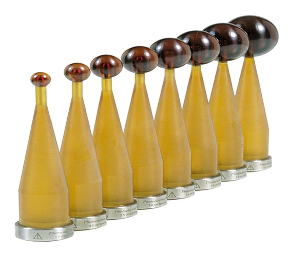
Intraoperative Radiotherapy (IORT) is a form of accelerated partial breast radiation that has been shown to be equivalent to conventional whole breast external beam radiotherapy (EBRT) in terms of local cancer control. However, questions have been raised about the potential of f IORT to produce breast parenchymal changes that could interfere with mammographic surveillance of cancer recurrence. The purpose of this study was to identify, quantify, and compare the mammographic findings of patients who received IORT and EBRT in a prospective, randomized controlled clinical trial of women with early stage invasive breast cancer undergoing breast conserving therapy between July 2005 and December 2009. Treatment groups were compared with regard to the 1, 2 and 4-year incidence of 6 post-operative mammographic findings: architectural distortion, skin thickening, skin retraction, calcifications, fat necrosis, and mass density. Blinded review of 90 sets of mammograms of 15 IORT and 16 EBRT patients demonstrated a higher incidence of fat necrosis among IORT recipients at years 1, 2, and 4. However, none of the subjects were judged to have suspicious mammogram findings and fat necrosis did not interfere with mammographic interpretation.
Figures


References
-
- Vaidya JS, Joseph DJ, Tobias JS, et al. Targeted intraoperative radiotherapy versus whole breast radiotherapy for breast cancer (TARGIT-A trial): an international, prospective, randomised, non-inferiority phase 3 trial. The Lancet. 2010;376(9735):91–102. - PubMed
-
- Luini A, Orecchia R, Gatti G, et al. The pilot trial on intraoperative radiotherapy with electrons (ELIOT): update on the results. Breast Cancer Research and Treatment. 2005;93(1):55–59. - PubMed
-
- Lemanski C, Azria D, Gourgon-Bourgade S, et al. Intraoperative radiotherapy in early-stage breast cancer: results of the montpellier phase II trial. International Journal of Radiation Oncology Biology Physics. 2010;76(3):698–703. - PubMed
-
- Elliott RL, Deland M, Head JF, Elliott MC. Accelerated partial breast irradiation: initial experience with the intrabeam system. Surgical Oncology. 2011;20(2):73–79. - PubMed
LinkOut - more resources
Full Text Sources
